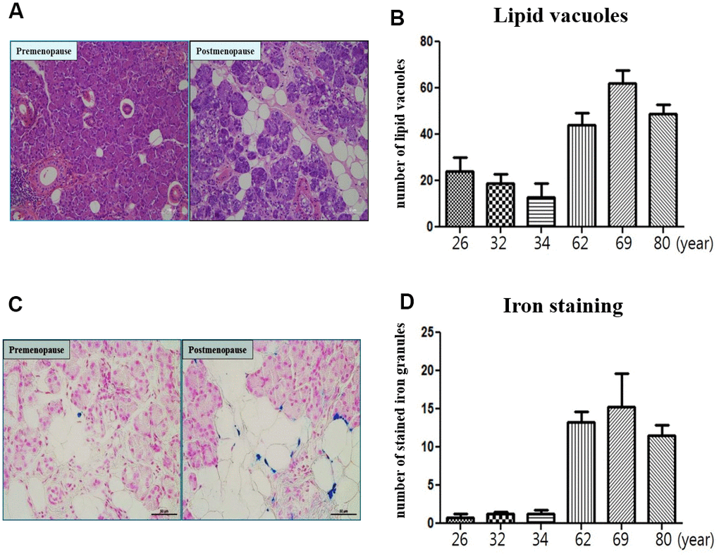
Figure 7.Microscopic finding of human submandibular gland tissue. (A) Lipid vacuoles of submandibular gland detected by H & E staining in the postmenopause group than premenopause group. (B) The number of lipid vacuoles of submandibular gland in the postmenopause group than premenopause group. (C) Iron accumulation of submandibular gland detected by Prussian Blue iron staining in the postmenopause group than premenopause group. (D) The number of stained iron of submandibular gland in the postmenopause group than premenopause group.