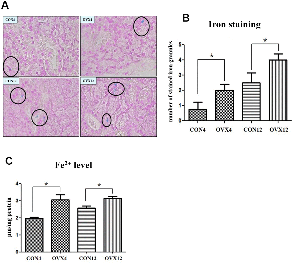
Figure 5.Iron deposition in submandibular gland. (A) The number of stained iron (black circle) detected by Prussian Blue iron staining. (B) Morphometric analysis of stained iron in the CON and OVX groups. (C) Cytosolic iron content in the CON and OVX groups. Two-way ANOVA test. *p<0.05, **p<0.01 and ***p<0.001. CON = control, OVX = ovariectomy.