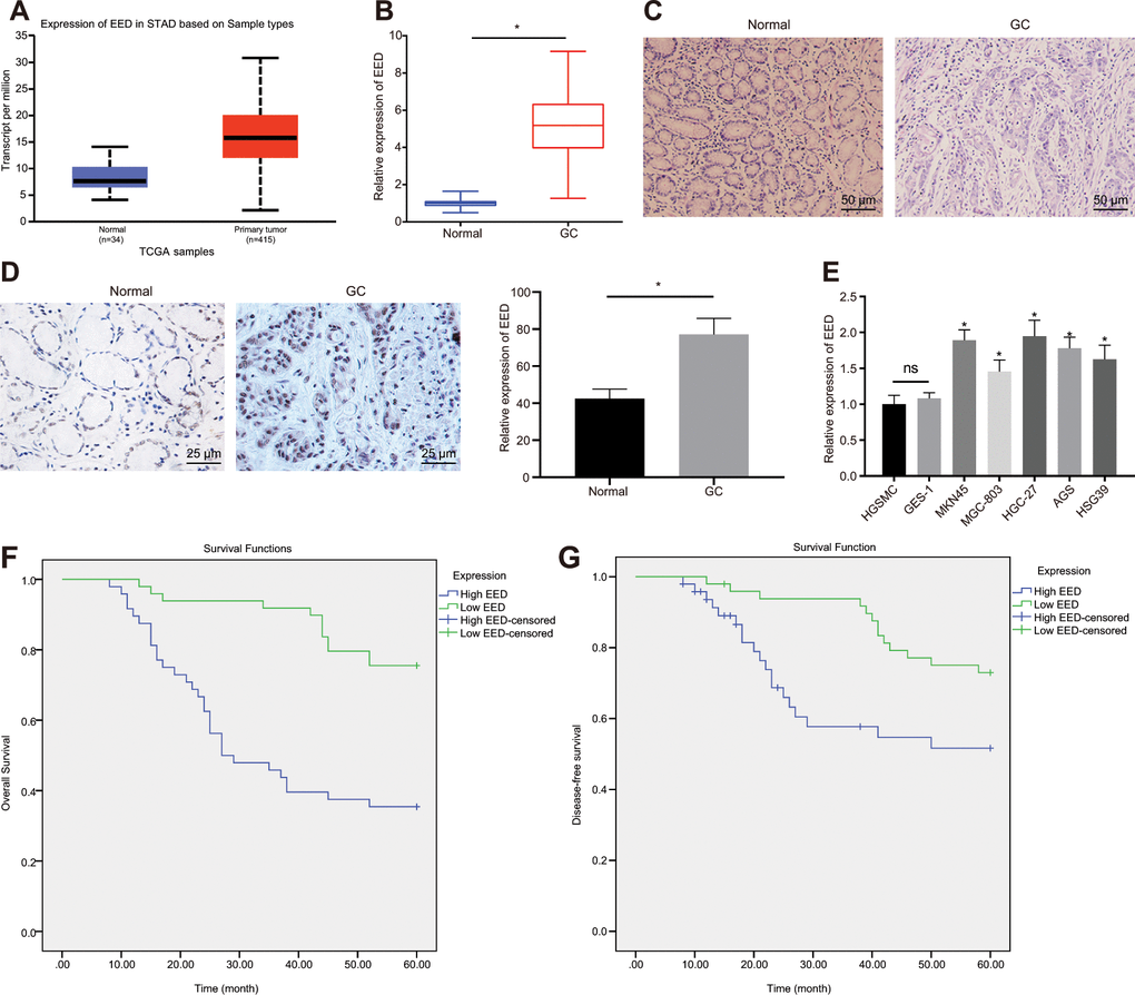
Figure 1.EED is highly expressed in GC tissues and cells. (A) Absolute EED expression in GC obtained by UALCAN database (http://ualcan.path.uab.edu/index.html). The blue box on the left represents the expression of normal samples, and the red box on the right represents the expression of GC samples. (B) RT-qPCR determination of EED expression in GC tissues and adjacent normal tissues (n = 97). (C) Representative images of GC tissues and adjacent normal tissues by HE staining. (D) Immunohistochemistry to assess the EED expression in GC tissues and adjacent normal tissues. (E) RT-qPCR to examine EED expression in GC cell lines and normal gastric cell lines, with β-actin as internal control. (F, G) Kaplan-Meier method with log-rank test to assess overall survival (F) and disease-free survival (G) of patients with relatively higher or lower EED expression (n = 97). Measurement data are expressed as mean ± standard deviation. * p < 0.05 compared with normal gastric cell line. GC tissues were compared with adjacent normal tissues by paired t test, n = 97. Data comparison among multiple groups was performed using one-way ANOVA with Tukey's post hoc test. Kaplan-Meier method was carried out to investigate the relationship between high and low expression of EDD in GC tissues and overall survival and disease-free survival (log-rank test). Cell experiments were repeated 3 times independently.
Figure 1 — Methylation of microRNA-338-5p by EED promotes METTL3-mediated translation of oncogene CDCP1 in gastric cancer | Aging