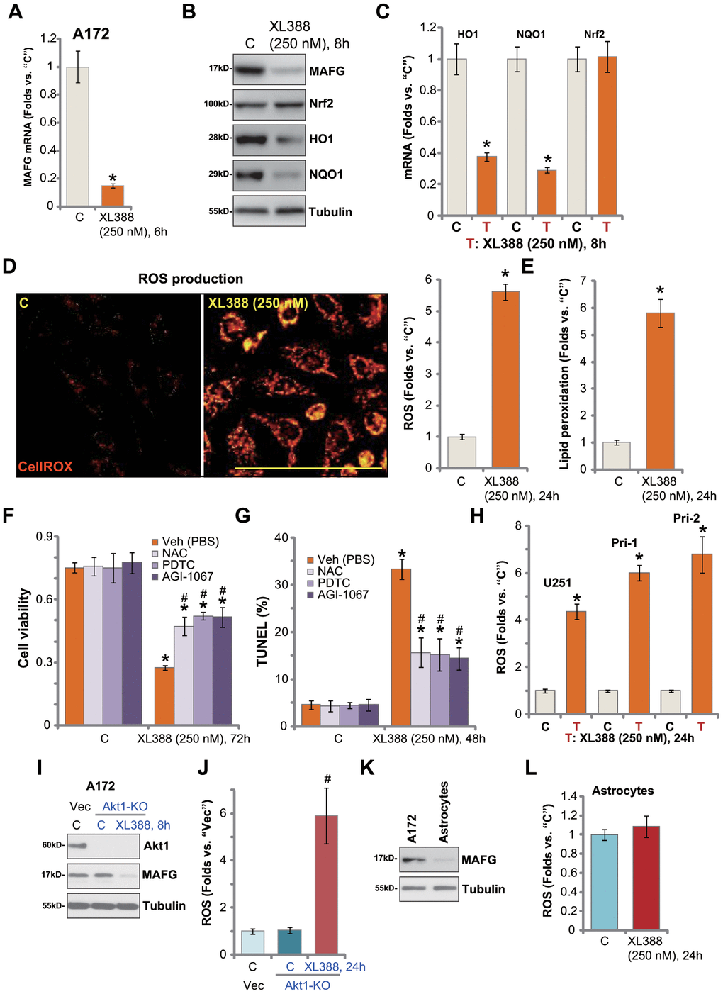
Figure 4.XL388 induces oxidative injury in human glioma cells. A172 cells or primary human glioma cells (“Pri-1”) were treated with XL388 (250 nM) and cultured for indicated time periods, then expression of listed mRNAs and proteins was tested by qPCR and Western blotting assays (A–C); Relative CellROX intensity (D) and lipid peroxidation (E) levels were tested. A172 cells were pretreated for 1h with n-acetylcysteine (NAC, 400 μM), pyrrolidine dithiocarbamate (PDTC, 10 μM) or AGI-1067 (10 μM), followed by XL388 (250 nM) stimulation for another 48-72h, then cell viability and apoptosis were tested by CCK-8 (F) and nuclear TUNEL staining (G) assays, respectively. U251MG (“U251”) and primary human glioma cells (“Pri-1/Pri-2”) were treated with XL388 (250 nM) for 12h, then the relative CellROX intensity was tested (H). A172 cells with the CRISPR/Cas9-Akt1-KO construct (“Akt1-KO” cells) or empty vector (“Vec”) were treated with or without XL388 (250 nM) and expression of listed proteins was shown (I). Relative ROS contents were tested by measuring CellROX intensity (J). Expression of MAFG protein in A172 cells and primary human astrocytes (“Astrocytes”) was shown (K); Astrocytes were treated with or without XL388 (250 nM) for 24h, and ROS intensity tested by CellROX assay (L). Data were presented as mean ± SD (n=5).* p <0.05 vs. “C” cells. #p <0.05. “Veh”-pretreated cells (F, G and J). Experiments in this figure were repeated three times, and similar results were obtained. Bar= 100 μm (D).