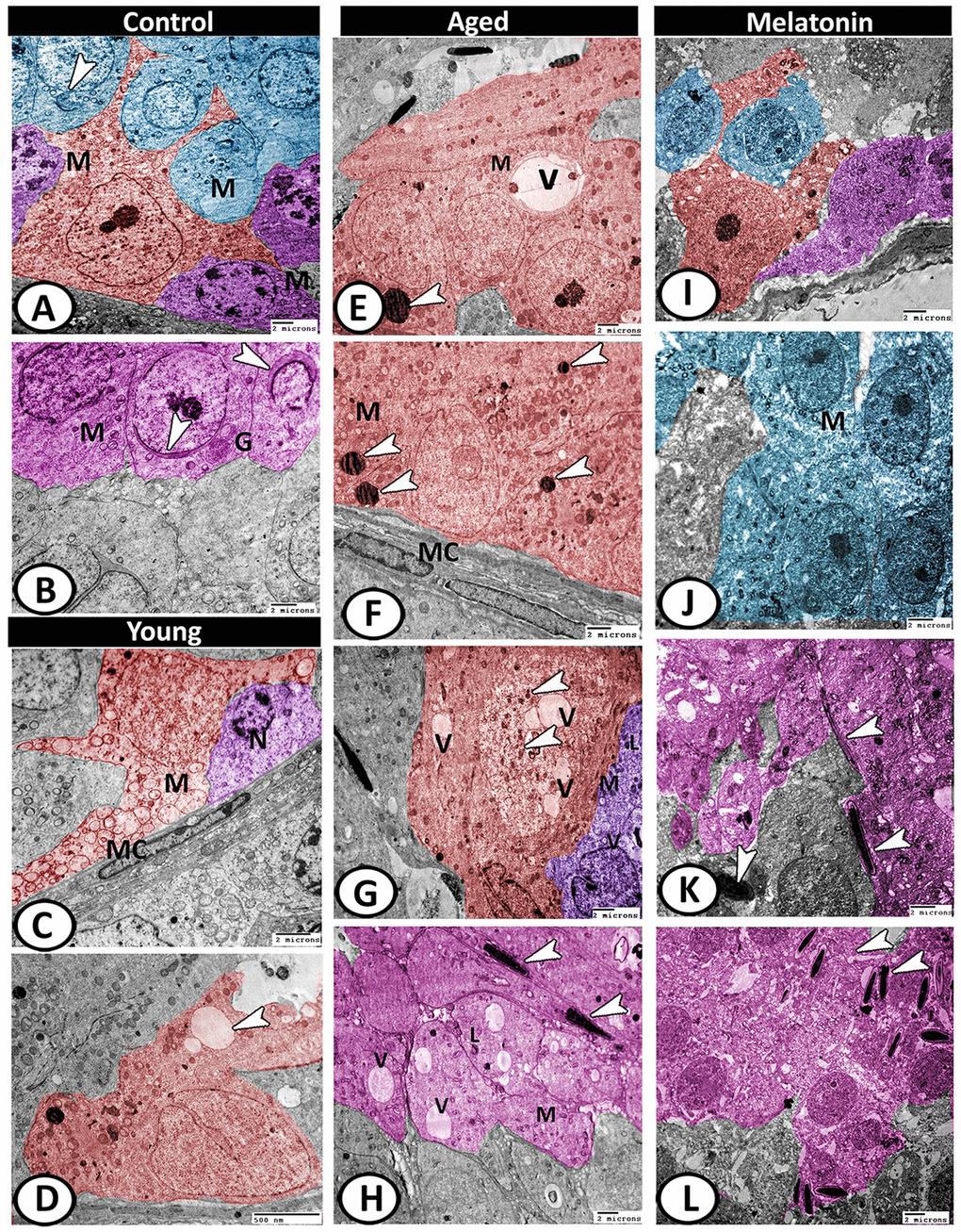
Figure 9.Digitally colored TEM image showing the ultrastructural changes in the seminiferous tubules of young and aged mice. (A) In the control group, the Sertoli cells (red) showed many cell processes and numerous mitochondria (M). Two spermatogonia populations (violet) were characterized by a network arrangement of nuclear chromatin and the cytoplasm contained mitochondria (M). The cytoplasm of primary spermatocytes (blue) was occupied by mitochondria (M)), and Golgi complex (arrowhead). (B) The spermatids (pink) characterized by the formation of an active acrosomal cap (arrowheads), presence of well-developed Golgi apparatus (G) and mitochondria (M). (C, D) In the young mutant mice, the Sertoli cells (red) showed degenerated mitochondria (M) and many vacuoles (arrowhead). The spermatogonia (violet) showed shrunken nuclei (N). Note, presence of myoid cells (MC) surrounding the seminiferous tubules. (E, F) In aged mice, the Sertoli cells' cytoplasm possessed vacuoles (V), mitochondrial metaplasia (M), lysosomes (arrowheads). Note, myoid cell (MC) showed no changes. (G) The cytoplasm of Sertoli cell (red) also contained phagocytosed materials (arrowheads) and vacuoles (V). The spermatogonia (violet) contained few mitochondria (M), vacuoles (V), and lysosomes (L). (H) The spermatids showed presence of many vacuoles (V), lysosomes (L), and the mitochondria (M) showed loss of their cristae. Many end pieces of degenerated sperms (arrowheads) could be demonstrated. (I) In melatonin group, the seminiferous tubules showed normal Sertoli cell (red), spermatogonia (violet) and spermatocytes (blue). (J) The spermatocytes (blue) contained numerous mitochondria (M) with some small vacuoles. (K, L) The differentiation of spermatids (pink) to normal sperms (arrowheads) was evident in melatonin group.