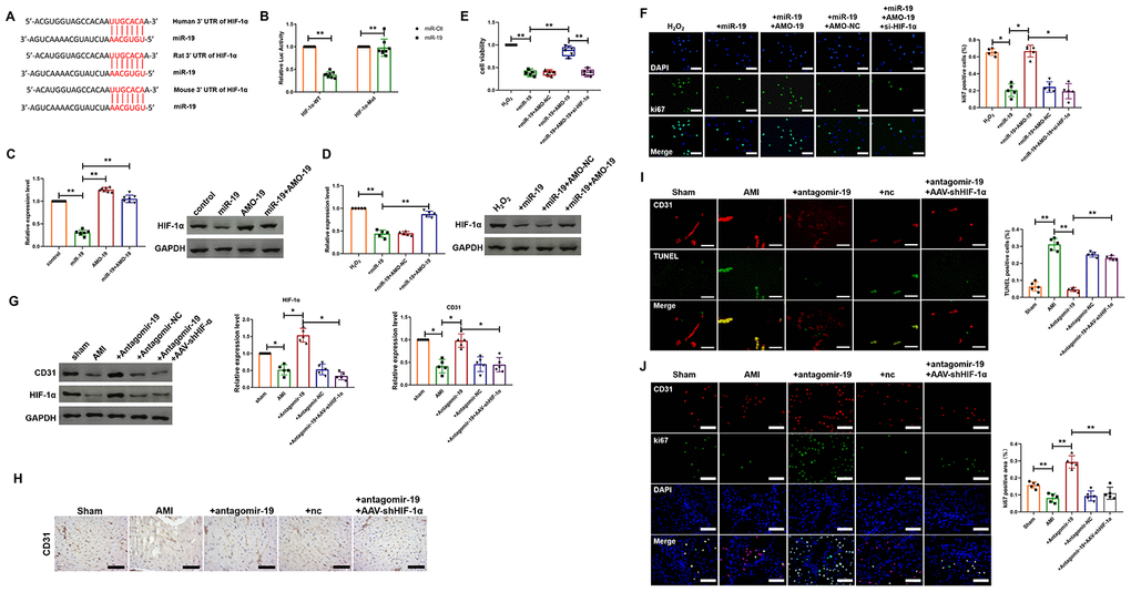
Figure 4.miR-19a-3p reduces endothelial cells proliferation and attenuates heart function by targeting HIF-1α. (A) The predicted binding site of miR-19a-3p on the 3’ UTR of HIF-1α gene. (B) Luciferase reporter activities of chimeric vectors carrying the luciferase gene and a fragment of the 3’ UTR of HIF-1α containing the wild type or mutant miR-19a-3p binding sites. (C) Western blot analysis of expression level of HIF-1α of endothelial cells transfected with miR-19a-3p or AMO-19. (D) Western blot analysis of expression level of HIF-1α of endothelial cells in response to H2O2 treatment with or without transfection of miR-19a-3p or AMO-19. (E) MTT assay of endothelial cells in response to H2O2 treatment with or without transfection of miR-19a-3p or AMO-19. (F) Immunofluorescence staining of ki67 in endothelial cells in response to H2O2 treatment with or without transfection of miR-19a-3p or AMO-19. (G) Western blot analysis of expression level of CD31 and HIF-1α in left ventricle of mice with or without MI. (H) Immunohistochemical staining of CD31 level in mice after MI. (I) TUNEL staining was performed to measure the level of endothelial cell death. (J) Immunofluorescence staining of ki67 in peri-infarct area of left ventricle. *P < 0.05 and **P < 0.01. All experiments were performed more than 4 biological repeats. Scar bar = 50μm.