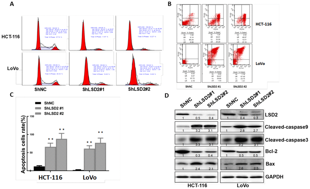
Figure 5.LSD2 promotes proliferation by inducing G1-S arrest and reducing apoptosis in colorectal cancer (CRC) cells. (A) HCT-116 and LoVo cells stably expressing vector or sh-LSD2#1 or sh- LSD2#2. The percentage of cells in G0/G1, S. or G2/M phases was tested using a subG1 assay and flow cytometry. (B) Apoptotic rates of HCT-116 and LoVo cells stably expressing vector or sh-LSD2#1 or sh-LSD2#2 measured by flow cytometry in annexin V/PI assays. LR, early apoptotic cells; UR, terminal apoptotic cells. Values represent the means ± SD. (C) Quantitative analysis of DNA apoptosis. Data were expressed as means ± SEM. **p < 0.01. (D) Bcl-2, BAX, CL-caspase 3, and CL-caspase 9 protein expression measured by Western blot with GAPDH protein as the loading control.