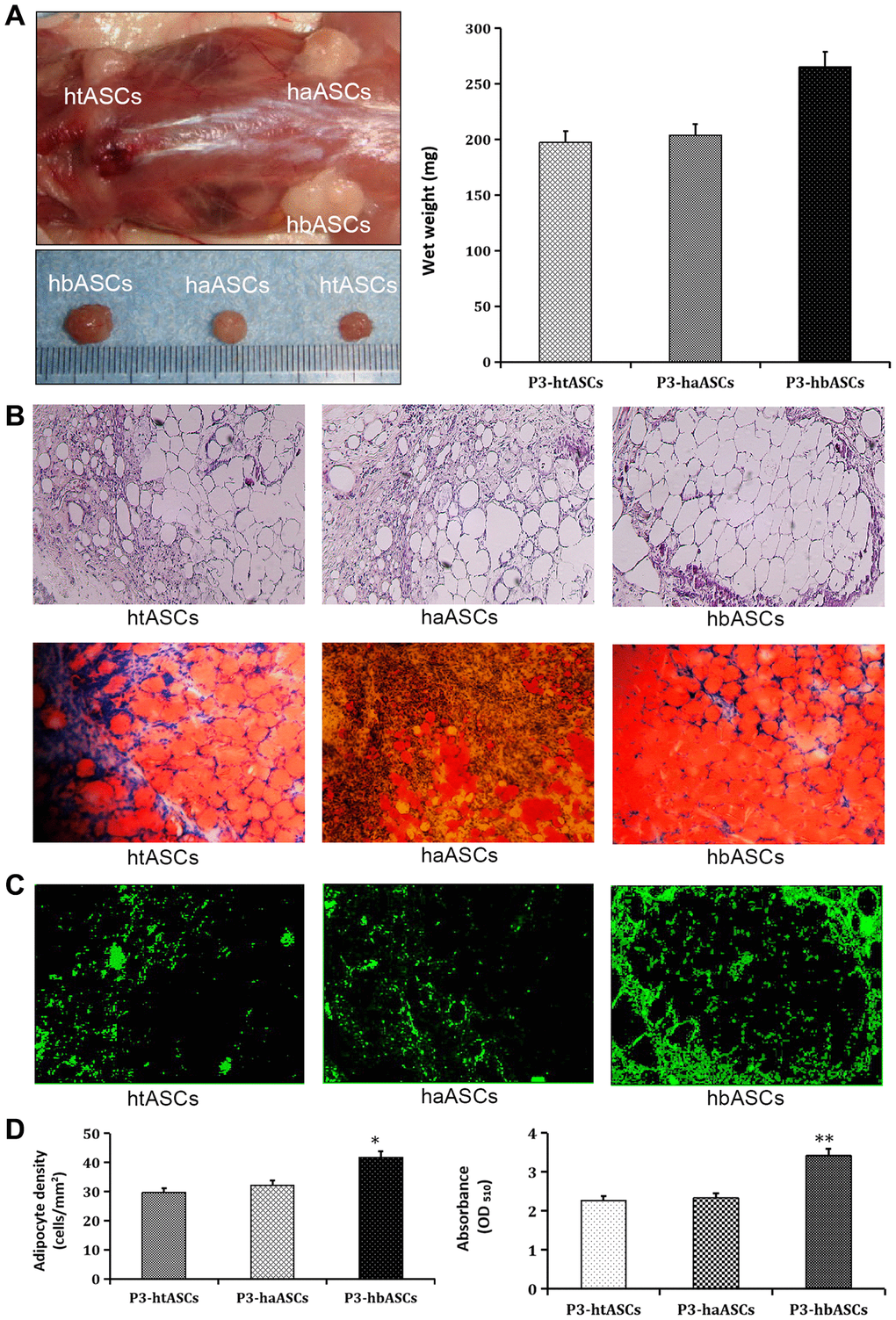
Figure 3.Different proliferation and differentiation abilities of haASCs, htASCs and hbASCs in vivo. (A) Regenerative adipose tissue macroscopic findings. Wet weights of regenerative adipose tissue in htASCs, haASCs, and hbASCs. *P<0.05. (B) H&E staining of the regenerative tissue after 12 weeks. The transplants derived from the three ASC types consisted predominantly of mature adipose tissue. Magnification, 100×. (C) GFP staining of the regenerative tissue after 12 weeks. In contrast to htASC or haASC tissue, GFP+ hbASC tissue contained larger Oil Red O-positive lipid droplets in the cytoplasm. GFP+ cells were detected in regenerative mature adipose tissue, indicating that these mature adipocytes had differentiated from GFP-labeled ASCs. Magnification, 100×. (D) Quantitative measurement of adipogenesis ability. Adipocyte density and intracellular lipid content were higher in hbASC tissue than in htASC or haASC tissue. *P < 0.05,**P < 0.05.