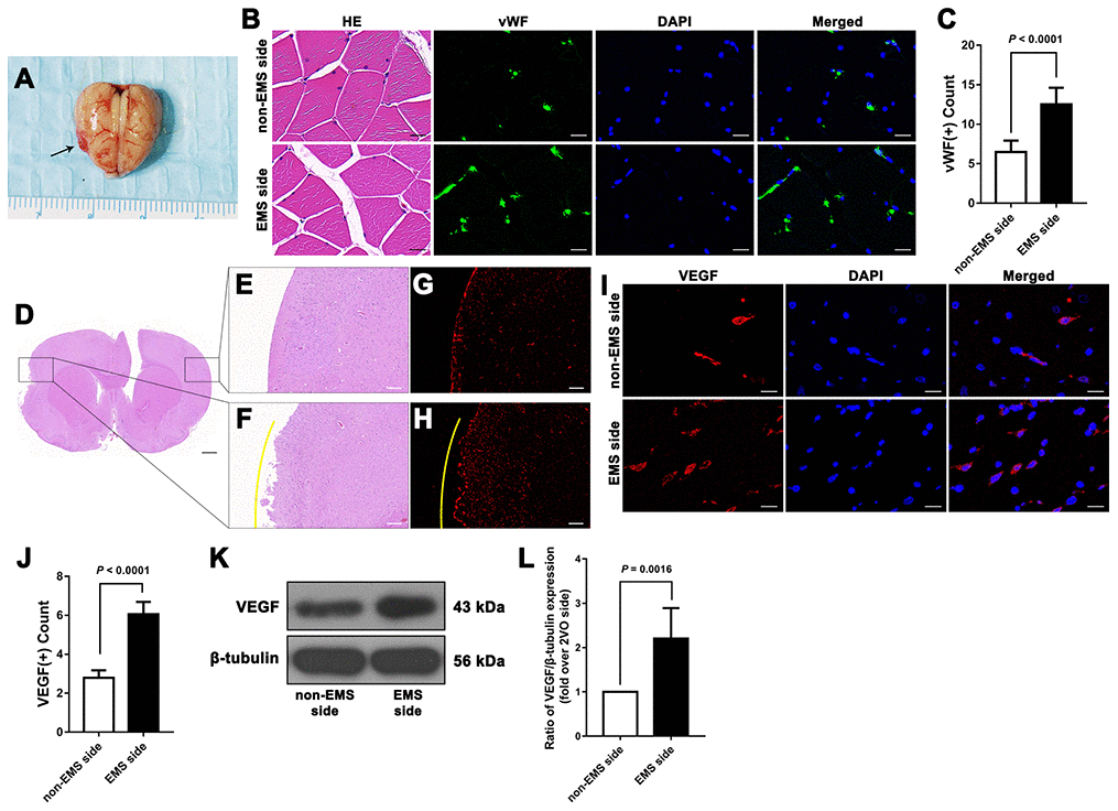
Figure 4.Effectiveness of the 2VO+EMS model in promoting EC proliferation determined by immunofluorescence and western blot assays. (A) Brain samples from 2VO+EMS rats (the black arrow indicates the adhered TM tissue). (B, C) Hematoxylin and eosin (HE) and immunofluorescence results showing that there was a significantly higher number of vWF(+) cells in the TM tissue on the EMS side than on the non-EMS side. Bar = 20 μm. (D) HE-stained slide of a brain from a 2VO+EMS rat. The two black frames indicate the portion of the brain tissue in contact with the TM and the brain tissue on the symmetrical part of the 2VO side. Bar = 1 mm. Enlarged image of the black frame on (E) the non-EMS side and (F) the EMS side. Bar = 200 μm. VEGF(+) immunofluorescence results for (G) the non-EMS side and (H) the EMS side. Bar = 200 μm. The yellow curve indicates the brain surface involved in EMS. Immunofluorescence (I, J) and western blot (K, L) results showing that there was significantly higher VEGF expression on the EMS side in the 2VO+EMS rat brains than on the non-EMS side. Bar = 20 μm. The error bars represent the ±SDs. VEGF: vascular endothelial growth factor; EMS: encephalo-myo-synangiosis; EC: endothelial cell; vWF: von Willebrand factor.