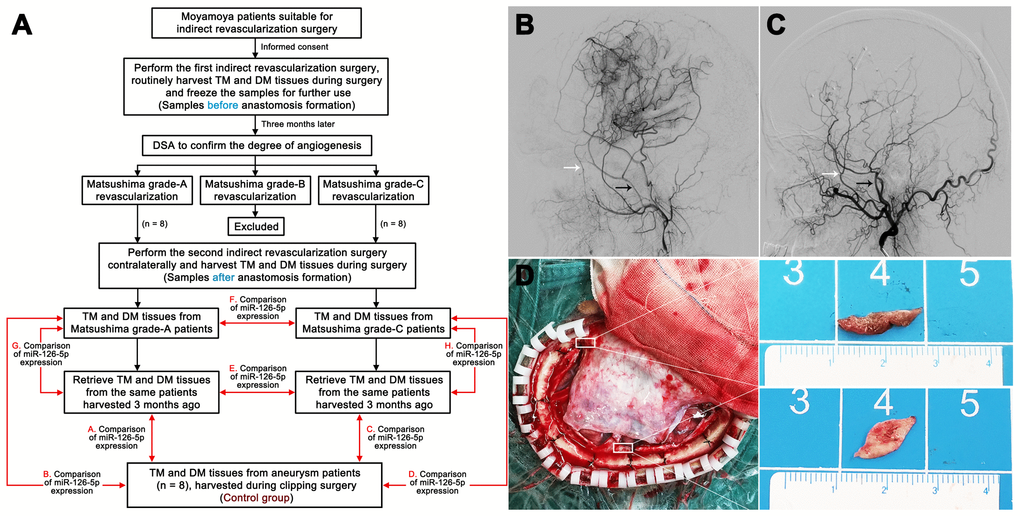
Figure 13.Harvesting of clinical samples. (A) Schedule of TM and DM sample harvesting. DSA films showing Matsushima grade-A revascularization (B) and Matsushima grade-C revascularization (C). The white arrows indicate the deep temporal artery, and the black arrows indicate the middle meningeal artery. (D) Intraoperative image showing the sources of the TM and DM samples. TM: temporal muscle; DM: dura mater.