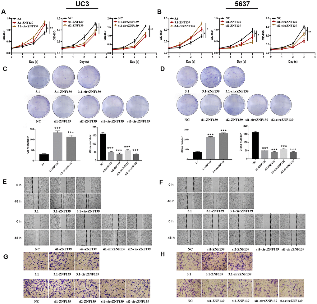
Figure 6.Cell proliferation, clone, migration, and invasion of UC3 and 5637 cells were evaluated after ZNF139/circZNF139 overexpression or knockdown. (A–B) CCK8 assay was employed to assess the proliferation of UC3 and 5637 cells with ZNF139/circZNF139 overexpression or knockdown. (C–D) Crystal violet staining was used to examine the colony formation of UC3 and 5637 cells with ZNF139/circZNF139 overexpression or knockdown. (E–F) Scratch wound healing assay was employed to evaluate the migration of UC3 and 5637 cells with ZNF139/circZNF139 overexpression or knockdown. Images of cell migration at 0 and 48 h transfection are shown at a magnification of 40×. (G–H) Transwell assay was used to analyze the invasion of UC3 and 5637 cells with ZNF139/circZNF139 overexpression or knockdown. Images are representative of the cells invading one field at a magnification of 100×. *, P<0.05; **, P<0.01. ZNF139, zinc finger with KRAB and SCAN domains 1; circ, circular; h, hour.