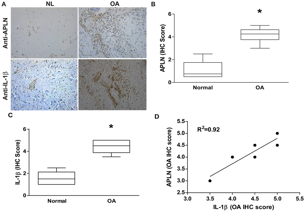
Figure 1.APLN expression is positively correlated with IL-1β expression in OA patients. (A) IHC staining showing increased levels of APLN and IL-1β expression in OA synovial tissue (n=8) compared to normal healthy tissue (n=5). (B, C) The IHC score of APLN and IL-1β are presented. (D) Correlation between levels of APLN and IL-1β expression in synovial tissues retrieved from OA patients.