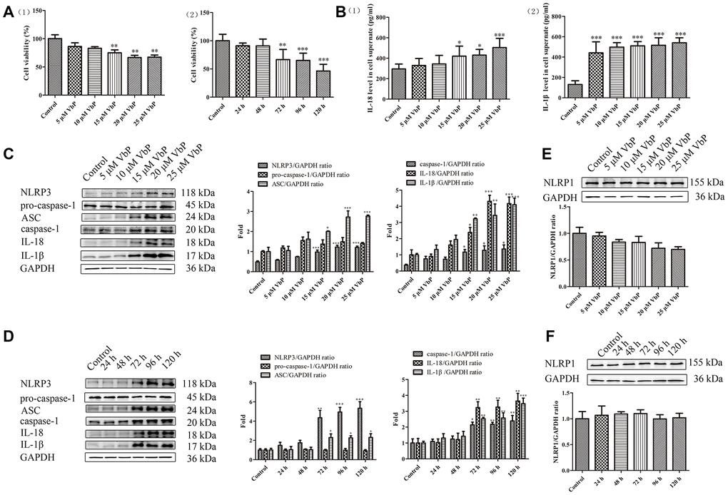
Figure 1.VbP induces pyroptosis in THP-1 macrophages. (A) Detection of cell viability (CCK-8 assay) after (1) 72 h exposure to different concentrations of VbP, and (2) exposure to 15 μM VbP for various times. (B) ELISA results showing secretion of IL-18 (1) and IL-1β (2) by macrophages treated for 72 h with different concentrations of VbP. (C) Dose-dependent expression of pyroptosis-related proteins in untreated (control) and VbP-treated macrophages (72 h exposure). (D) Time-dependent expression of pyroptosis-related proteins in untreated (control) and VbP-treated (15 μM) macrophages. (E) Dose-response analysis of NLRP1 inflammasome expression in VbP-treated macrophages (72 h exposure). (F) Time-response analysis of NLRP1 expression in macrophages exposed to 15 μM VbP. n = 3; *P<0.05, **P<0.01, and ***P<0.001 vs. control cells.