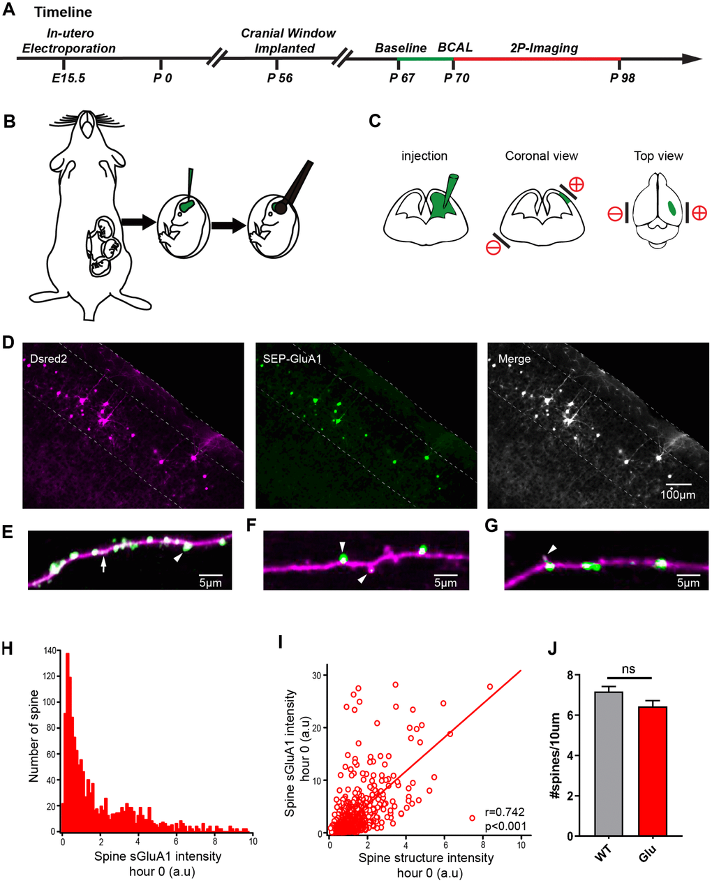
Figure 4.Expression of SEP-GluA1 in layer 2/3 somatosensory cortex neurons in vivo. (A) Timeline of experimental design. The green line and the red line indicates the two-photon imaging time course. (B, C) Schematic drawing of in utero electroporation. +/- means positive and negative polar, respectively. (D) Representative images showing expression of dsRed2 (purple), SEP-GluA1 (green) and their overlap (white). (E–G) SEP-GluA1 in green, dsRed2 in magenta, and their overlap in white. (H) Histogram of spine sGluA1 intensity before BCAL at hour 0. (I) Correlation between spine sGluA1 and spine size before BCAL at hour 0. n= 1381 spines. r, Pearson's linear correlation coefficient, p value is from Monte-Carlo shuffling. (J) Quantification of spine density. n= 27 neurons in 5 control mice, 24 neurons in 5 Glu mice. ns, not significant, Student’s t-test. Error bars = s.e.m.