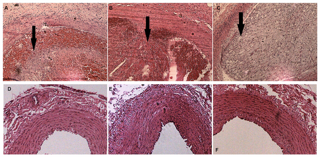
Figure 2.H&E staining of rabbit carotid arteries. (A–C) H&E-stained vasculature shows plaques of varying degrees in rabbit carotid arteries in the AS group as the arrows show, ×40. (D–F) H&E-stained arteries show that no evident abnormal changes are detected in rabbit carotid arteries of the case group, ×40.