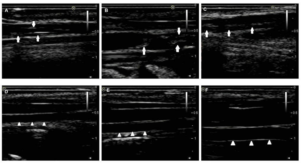
Figure 1.2D-ultrasound images of rabbit carotid arteries at the 12th week. (A–C) 2D-ultrasound images reveal that obvious atherosclerotic plaques formed on rabbit carotid arterial intima as the arrows show. (D–F) 2D-ultrasound images demonstrate that carotid arteries intima of rabbits treated with normal chow still maintain smoothness as the arrows show.