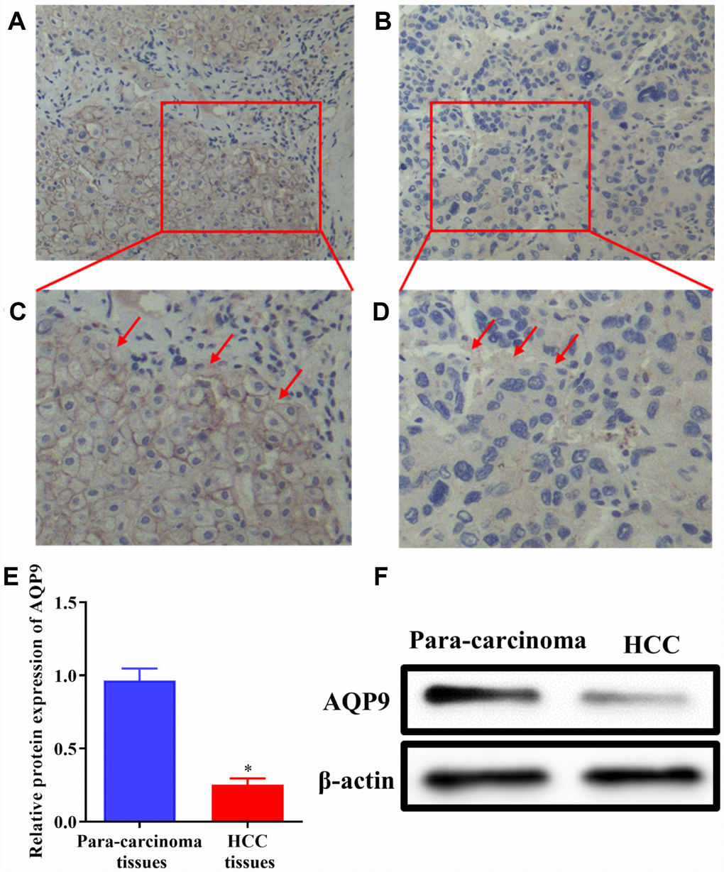
Figure 1.The expression of AQP9 was reduced in HCC samples. (A–D) The levels of AQP9 were examined in HCC and paired non-tumor tissues by immunohistochemistry analysis (magnificationx200 and x400). (E and F) The levels of AQP9 were also examined using western blotting. The results were represented as mean ± SD. P<0.05 vs. para-carcinoma control. Each experiment was repeated 3 times. AQP, aquaporin 9; HCC, hepatocellular carcinoma.