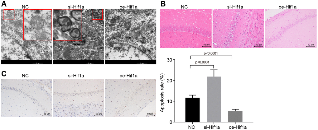
Figure 5.The pathological changes of hippocampal neurons in the MPTP-lesioned mouse model of PD. (A) The ultrastructure of hippocampal neurons treated with si-Hif1a or oe-Hif1a to electron microscopy. (B) The pathological changes of hippocampal neurons treated with si-Hif1a or oe-Hif1a revealed by HE staining (scale bar = 50 μm). (C) The apoptosis of hippocampal neurons treated with si-Hif1a or oe-Hif1a detected by TUNEL staining (scale bar = 50 μm). * p < 0.05 vs. the NC group (MPTP-lesioned mice treated with NC plasmids). N = 10.