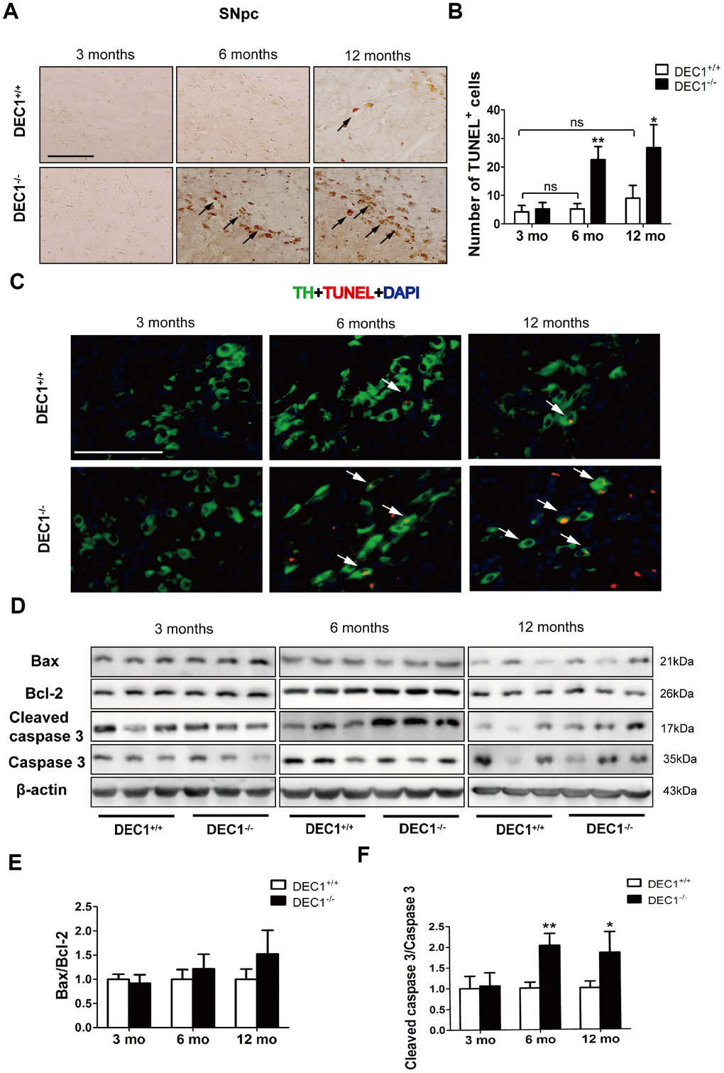
Figure 4.DEC1 deficient mice show an increase in neuron apoptosis in the SNpc. (A) Representative images of TUNEL+ cells in the SNpc of DEC1+/+ and DEC1-/- mice at 3, 6 and 12 months(n=4 in each group). TUNEL+ apoptotic cells were labeled by black arrowheads. (B) The number of TUNEL+ cells (Two-way AONVA, gene: F(1,18)=42.961, p<0.01; age: F(2,18)=18.002, p<0.001; interaction: F(2,18)=9.031, p=0.002). (C) Representative images of TH (red), TUNEL (green) and DAPI(blue) in the SNpc of DEC1+/+ and DEC1-/- mice at the age of 3, 6 and 12 months(n=4 in each group). TUNEL+ apoptotic cells co-expressed with TH+ cells were labeled by white arrowheads. (D) The expression of the apoptosis-related proteins at the age of 3, 6 and 12 months (n=6 in each group). (E) Bax/Bcl-2 (Two-way AONVA, gene: F(1,18)=4.743, p=0.069; age: F(2,18)=2.419, p=0.117; interaction: F(2,18)=2.419, p=0.117). (F) The cleaved caspase 3/caspase 3 (Two-way AONVA, gene: F(1,18)=25.945, p<0.01; age: F(2,18)=5.518, p=0.014; interaction: F(2,18)=5.518, p=0.014). The data are analyzed using t-test for the same age in two genotypes of mice and expressed as mean ± SD. *p<0.05, **p<0.01 vs the age-matched DEC1+/+ mice. ns p>0.05, comparisons are shown in the figure. Scale bar=100 μm.