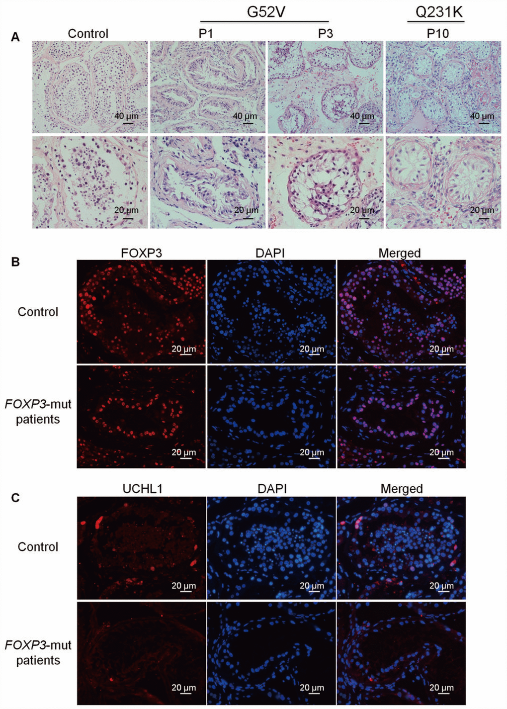
Figure 2.Morphology and phenotype of FOXP3-mut NOA patients and OA controls. (A) H&E staining revealed that seminiferous tubule diameter was reduced and spermatogenesis was arrested in FOXP3-mut NOA patients. Scale bars = 40 μm and 20 μm, respectively. (B-C) Immunohistochemical staining demonstrated the expression of FOXP3 protein (B) and UCHL1 protein (C) in FOXP3-mut NOA patients (low panels) and OA controls (upper panels). Experiments were repeated for at least three times. Scale bars = 20 μm.