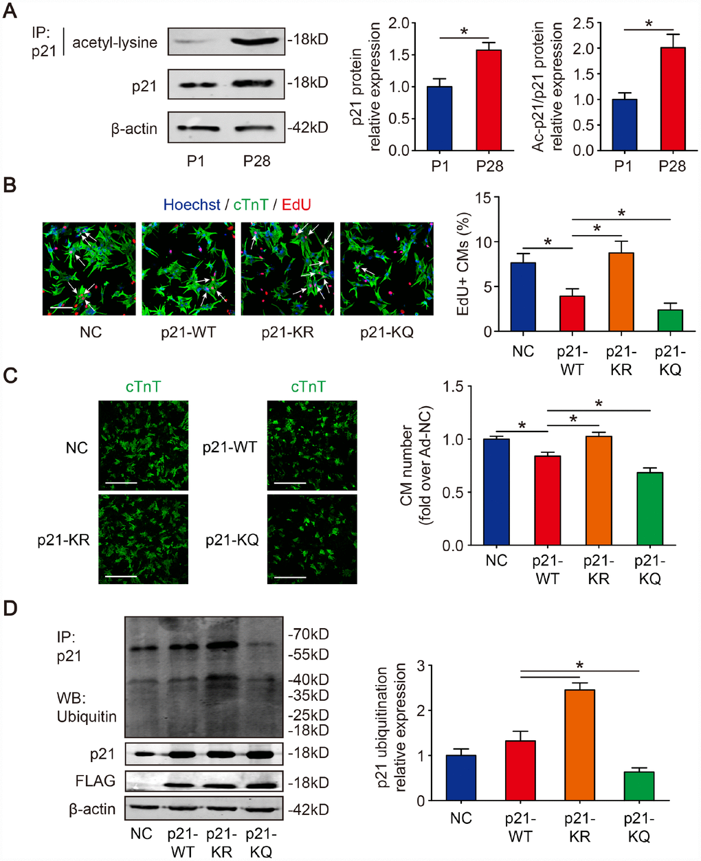
Figure 1.Deacetylation of p21 attenuates p21 stability and CM cell cycle arrest. (A) P1 and P28 mouse heart lysates were immunoprecipitated with a p21 antibody and analyzed by Western blotting with acetyl-lysine antibody; Western blotting was used to evaluate p21 protein expression in P1 and P28 mouse heart lysates. β-actin was used as a loading control (n=5). (B) Isolated P1 CMs were transfected with NC, p21-WT, p21-KR, or p21-KQ. EdU incorporation was detected by immunofluorescence. Scale bar, 50 μm. Quantitative analyses represent fields from 5 mice per group. (C) Isolated P1 CMs were transfected with NC, p21-WT, p21-KR, or p21-KQ and quantification of counts from P1 CMs transfected with NC, p21-WT, p21-KR, or p21-KQ (n=5). cTnT immunofluorescence was assessed to detect CM numbers. Scale bar, 500 μm. (D) Isolated P1 CMs were transfected with NC, FLAG-p21-WT, FLAG-p21-KR or FLAG-p21-KQ. Whole cell lysates were immunoprecipitated with a p21 antibody and analyzed by Western blotting with an ubiquitin antibodies; Western blotting was used to detect p21 levels. β-actin was used as a loading control (n=5). Statistical significance was calculated using a two-tailed unpaired Student’s t-test in (A) and a one-way ANOVA followed by an LSD post hoc test in (B–D). *p<0.05; data are presented as the mean ± S.E.M.