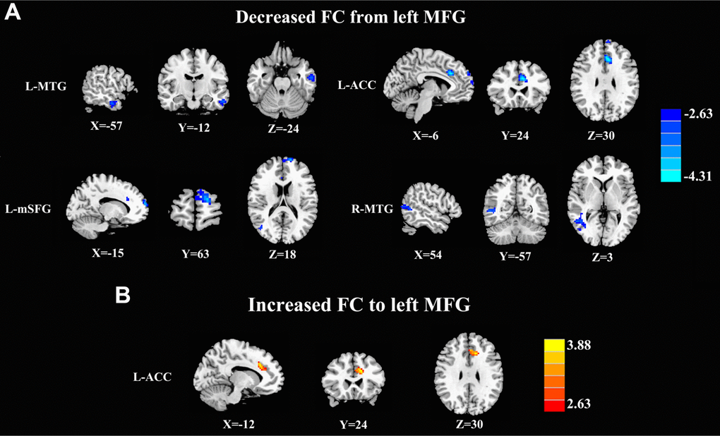
Figure 2.Aberrant causal connectivity from the bilateral SFG in mTBI patients at the acute stage. (A) Decreased causal connectivity from the left MFG to the L-MTG, R-MTG, L-ACC, and L-mSFG. (B) Increased causal connectivity from the left MFG to the left ACC. L, left; R, right; MFG, middle frontal gyrus; MTG, middle temporal gyrus; ACC, anterior cingulate cortex; mSFG, medial superior frontal gyrus.