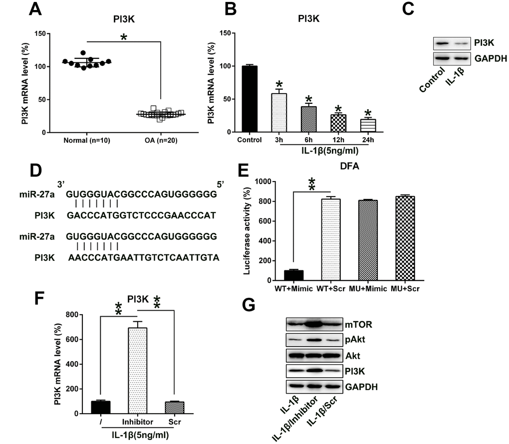
Figure 6.MiR-27a directly targets PI3K. (A) PI3K expression in normal (n=10) as well as OA cartilage samples (n=20) were evaluated via Q-PCR. (B, C) Chondrocytes were treated with IL-1β (5 ng/ml) for 0, 6, 12, or 24 hours. The PI3K expression was assessed by Q-PCR and WB analyses. (D) Graphical representation of the conserved miR-27a-binding motif at the 3′-UTR of PI3K. (E) The DLRA was performed following the transfection of the cells with the luciferase reporter constructs that included either mutated (MU) or wild-type (WT) sequences of human PI3K 3'-UTR after transfection with the miR-27a mimic/miR-Scr. The luciferase activity was normalized to the β-galactosidase activity. Q-PCR (F) as well as WB (G) analyses were performed to assess the PI3K transcription and translation levels, as well as the Akt, phosphor-Akt, and mTOR protein levels, following the transfection of IL-1β-treated chondrocytes transfected with the miR-27a inhibitor and miR-Scr. The results are described as the mean ± SD. *P < 0.05, **P < 0.05 vs. indicated group.