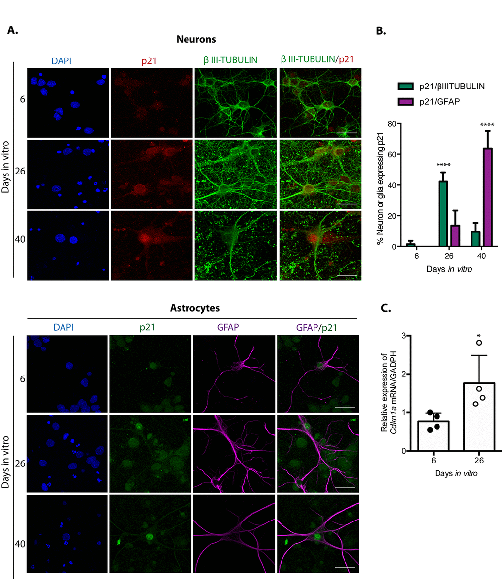
Figure 2.Neuronal cells in cortical long-term culture showed increased expression of p21CIP1/WAF1. (A) Immunofluorescence to detect p21CIP1/WAF1 (p21) in neurons (expressing βIII-TUBULIN) or astrocytes (expressing GFAP) in primary culture of cortical cells incubated during the indicated DIV. Notice that mostly neurons increased the abundance of p21CIP1/WAF1 at 26 DIV, indicating that neurons acquired senescent features before glial cells. Scale bar represents 25 μm. Arrows indicate examples of cells with healthy nuclei counted (not all the healthy cells are indicated). (B) Percentage of neurons or glial cells expressing p21CIP1/WAF1 over all cells. The mean of three independent experiments, each done by duplicate, is plotted. Bars represent standard deviation. Two-way RM ANOVA analysis, with Tukey´s multiple comparison test. **** p<0.0001. (C) qRT-PCR from total RNA purified from cortical primary cultures during the indicated days. The relative expression of Cdkn1a mRNA was normalized with Gapdh mRNA. Bars represent SD. * p=0.039 by unpaired t test two tailed. n=4.