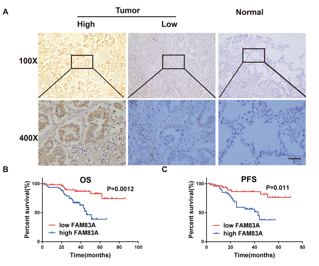
Figure 2.FAM83A was highly expressed in NSCLC tissues and correlated with worse survival. (A) Immunohistochemical staining showed that FAM83A was highly expressed in NSCLC tumors (n = 49/101) compared with normal lung tissues (n = 2/50, P < 0.05). (B–C) Kaplan-Meier plots of overall-survival (B) and progression-free survival (C) in NSCLC patients with high and low levels of FAM83A. Scale bar, 100 μm.