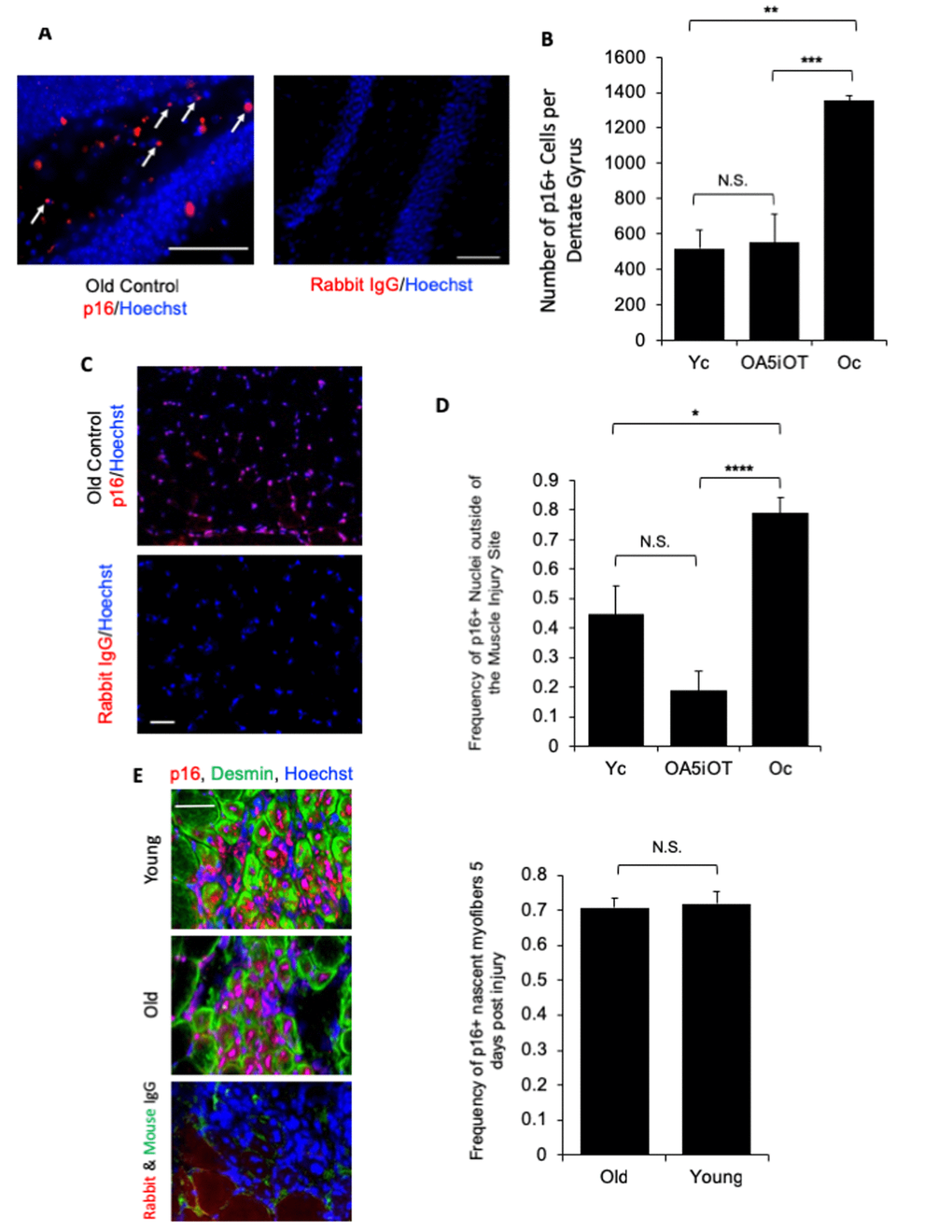
Figure 5.p16 levels are decreased in situ in muscle and brain by Alk5i+OT. Cryosections of injured and uninjured muscle (10 µm, each) and brain (25 µm) were assayed for the number of p16+ nuclei by immunofluorescence, using Hoechst to label all nuclei. p16 showed the predicted nuclear localization in these assays. (A) Representative images of p16+ cells in the polymorphic layer near the dentate gyrus in an old control brain and isotype-matched IgG non-specific immunofluorescence are shown. Arrows point to p16+ (red)/Hoechst+ (blue) nuclei in the stated region. (B) The number of p16+ cells in the polymorphic layer of the hippocampus was decreased by the Alk5i+OT treatment, ***old control & Alk5i+OT p=0.0003, scale bar=50 µm at 40x magnification, IgG scale bar=50 µm at 20x magnification. Young control (Yc) n=5, old control (Oc) n=8, Alk5i+OT (OA5iOT) n=8. (C) Representative images of p16+ nuclei outside of the injury site in the TA muscle of an old vehicle control mouse at 5 days post single CTX injection and isotype-matched IgG non- specific immunofluorescence, are shown; scale bar=50 µm at 20x magnification. (D) The number of p16+/Hoechst+ nuclei divided by the total number of nuclei (Hoechst+) per field-of-view at 20x magnification (frequency of p16+ nuclei) was quantified. The frequency of p16+ nuclei outside of injury sites is significantly greater in muscle of old vehicle-treated control mice, as compared to young control and old Alk5i+OT treated. N=5 for each cohort. Young control (Yc), old+Alk5i+OT (OA5iOT), old control (Oc) N.S. = P-value Yc &OA5iOT = 0.064, *** = P-value Yc & Oc = 0.011, **** = P-value OA5iOT & Oc = 0.000064. (E) Representative images of the sites of injury/regeneration of young and old TA muscle from control - HBSS treated mice; 10-micron sections desmin (green), p16 (red) immunofluorescence and isotype-matched IgG control non-specific immune-fluorescence, are shown. Scale bar is 50 micron at 40x magnification. Robust p16+ nuclei are observed in both young and old muscle, and many of these are in centrally-nucleated newly formed desminhigh myofibers. The frequency of p16+ centrally-nucleated myofibers was quantified (right). The relative number of these p16+ fibers were found to be nearly identical in young injured (n=5) and old injured (n=7) muscle. N.S. = P-value Old and Young = 0.7918.