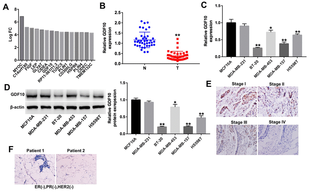
Figure 2.GDF10 expression in human TNBC specimens and cell lines. (A) Differentially expressed mRNAs (log2 fold-change (FC) ≥ 4) in TNBC samples. (B) Relative expression of GDF10 in tumors and adjacent normal tissues (N) from TNBC patients (n = 40). **P < 0.01, compared with the N group. (C) The levels of GDF10 in TNBC cell lines (MDA-MB-231, BT-20, MDA-MB-453, MDA-MB-157 and HS598T), and in normal mammary epithelial cells (MCF10A) were detected by qRT-PCR. *P < 0.05, **P < 0.01, compared with MCF10A cells. (D) GDF10 expression in MCF10A, MDA-MB-231, BT-20, MDA-MB-453, MDA-MB-157 and HS598T cells assessed by western blotting. β-actin was used as internal control. *P < 0.05, **P < 0.01, compared with MCF10A cells. (E) Representative GDF10 IHC staining (×200) of stages I, II, III, and IV TNBC samples. (F) IHC staining images of ER-negative nuclear expression, PR negative nuclear expression and HER-2/neu negative expression (magnification x 200) in patients with TNBC.