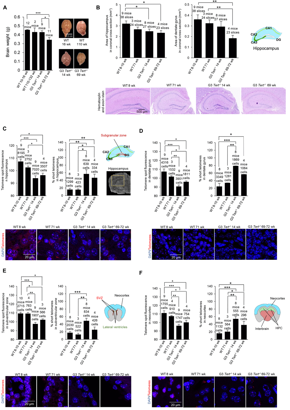
Figure 1.Mice deficient for telomerase have smaller brains, shorter telomeres, more proliferation, more DNA damage, and less neurogenesis. (A) Brain weight and representative images of young and old wild-type and G3 Tert-/- mice. (B) Area of hippocampus and dentate gyrus in untreated mice quantified from representative images of brain sections stained with hematoxylin and eosin. (C) Q-FISH for telomere spot fluorescence measured in the hippocampus, (D) the dentate gyrus specifically, (E) the subventricular zone, and (F) the neocortex. The mean telomere spot fluorescence is shown. The percentage of short telomeres is also shown with “short” being defined as a fluorescence intensity less than the 15th percentile of the fluorescence intensity values of a control sample. Cartoon diagrams label the different regions of the brain. In part (C), A scan of a coronal brain cross-section without fluorescence is shown with the hippocampus region highlighted in yellow. Representative images show the telomere spots labeled with Cy3-Tel probe (in red), and nuclei stained with DAPI (blue).