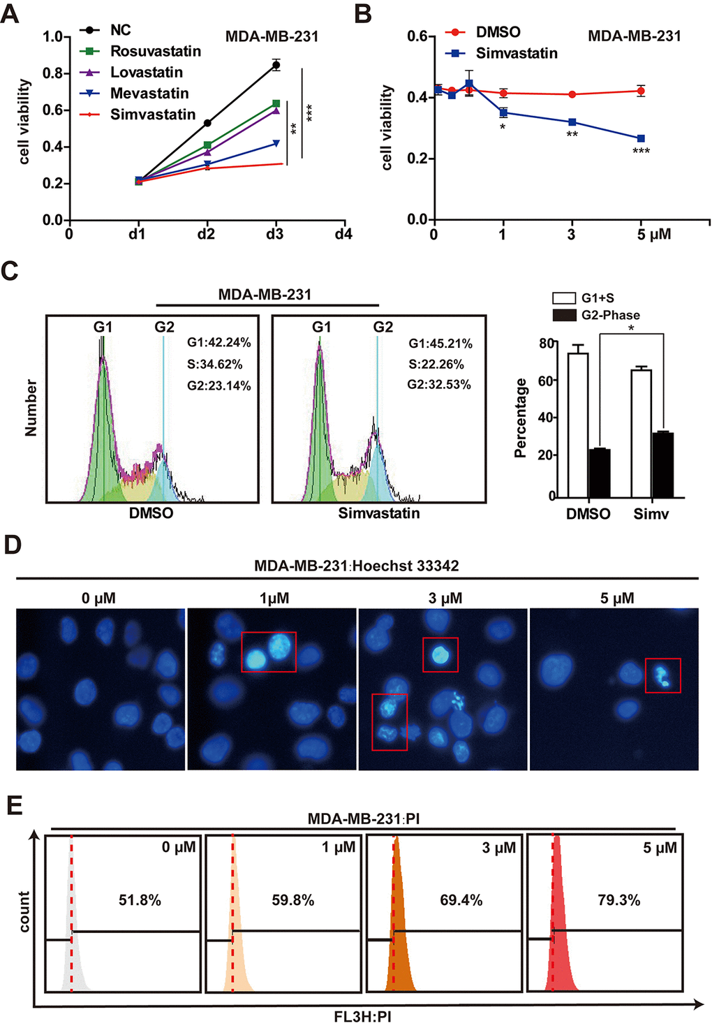
Figure 1.Effects of simvastatin on cell proliferation and cell death in MDA-MB-231 cells. (A) MDA-MB-231 cells were treated with 1µM rosuvastatin, lovastatin, mevastatin or simvastatin for 24h and 48h. (B) MDA-MB-231 cells were treated with varying concentrations (1-5µM) of simvastatin for a period of 48h. All the cell viability (cell proliferation) assays were analyzed by the CCK-8 assay. (C) the effects of simvastatin on the cell cycle were measured by flow cytometry with PI staining. (D) Representative photomicrography of treated MDA-MB-231 cells with varying doses simvastatin showing nuclei fragmentation. (E) MDA-MB-231 were treated with various doses of simvastatin for 48h. Cell death was determined by PI FACS analyses. The percentage of necrotic/apoptosis cells (PI positive) were moved to the right quadrant (Relative to 0µM). The p-values were calculated using standard Student t-tests. Error bars represent mean± SEM of three individual experiments. *** P ≤ 0.001, ** P ≤ 0.01, * P ≤ 0.05.