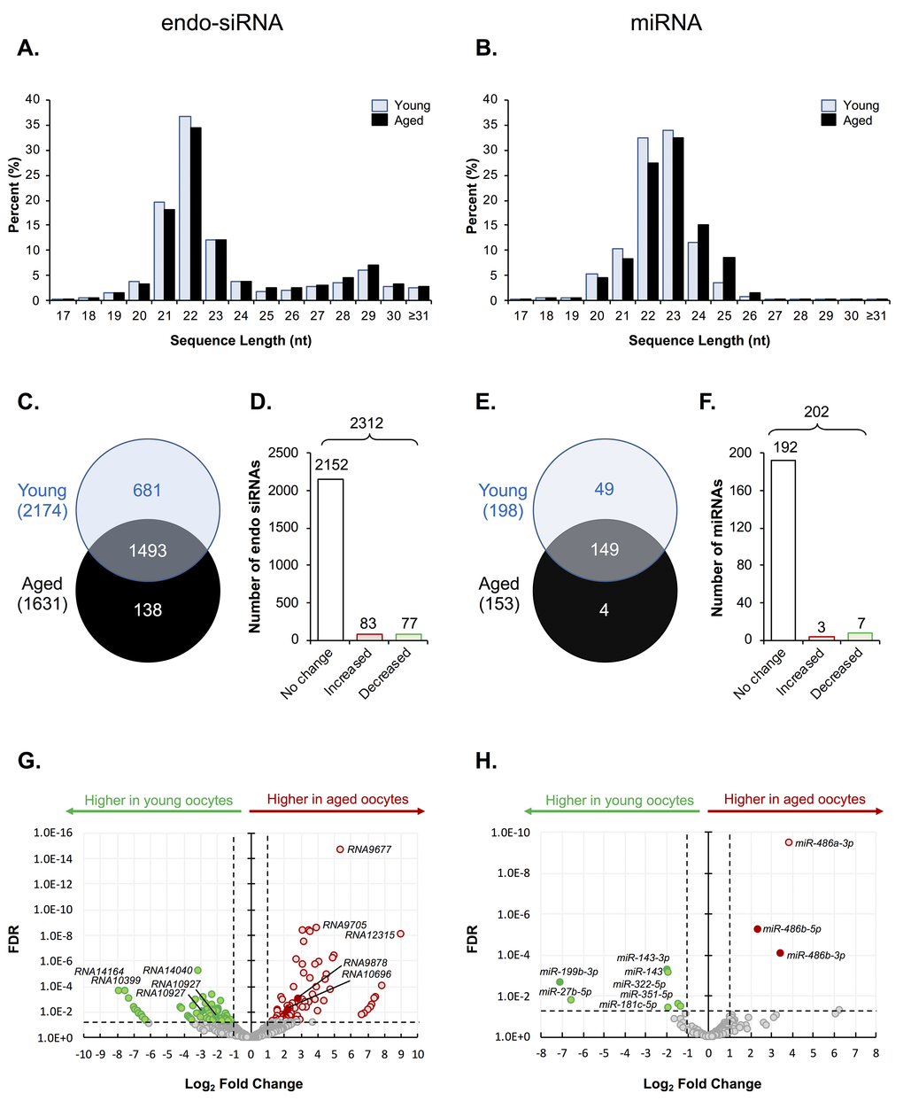
Figure 1.Endo-siRNA and miRNA signatures of young and aged oocytes. Following filtering and normalization, sRNA reads were mapped to known endo-siRNA and miRNA from RNAcentral sequence database (August 2017, RNAcentral) (https://rnacentral.org/) to explore changes in the endo-siRNA and miRNA landscape between young and aged oocytes. (A) Endo-siRNA and (B) miRNA sequence length distribution between young and aged GV oocytes. (C) Venn diagram illustrating the total number of endo-siRNA and (E) miRNA identified in young and aged oocytes. (D) Graphical representation of the proportion of endo-siRNAs and (F) miRNAs, identified as being expressed at equivalent levels (unchanged) or that were up- or down-regulated (increased or decreased, respectively) between young and aged oocytes. (G) Volcano plots depicting log2-fold changes (x-axis) and false discovery rate (FDR; y-axis) of endo-siRNAs and (H) miRNAs between young and aged oocytes. Solid dots represent sRNAs that were selected for RT-qPCR validation. Counts of ≥ 10 reads aligning to a specific sRNA was used as a threshold for a positive endo-siRNA or miRNA identification in this study. sRNAs experiencing a threshold of ≥ ± 2-fold change and false discovery rate of < 0.05 were considered as being differently expressed between young and aged oocytes.