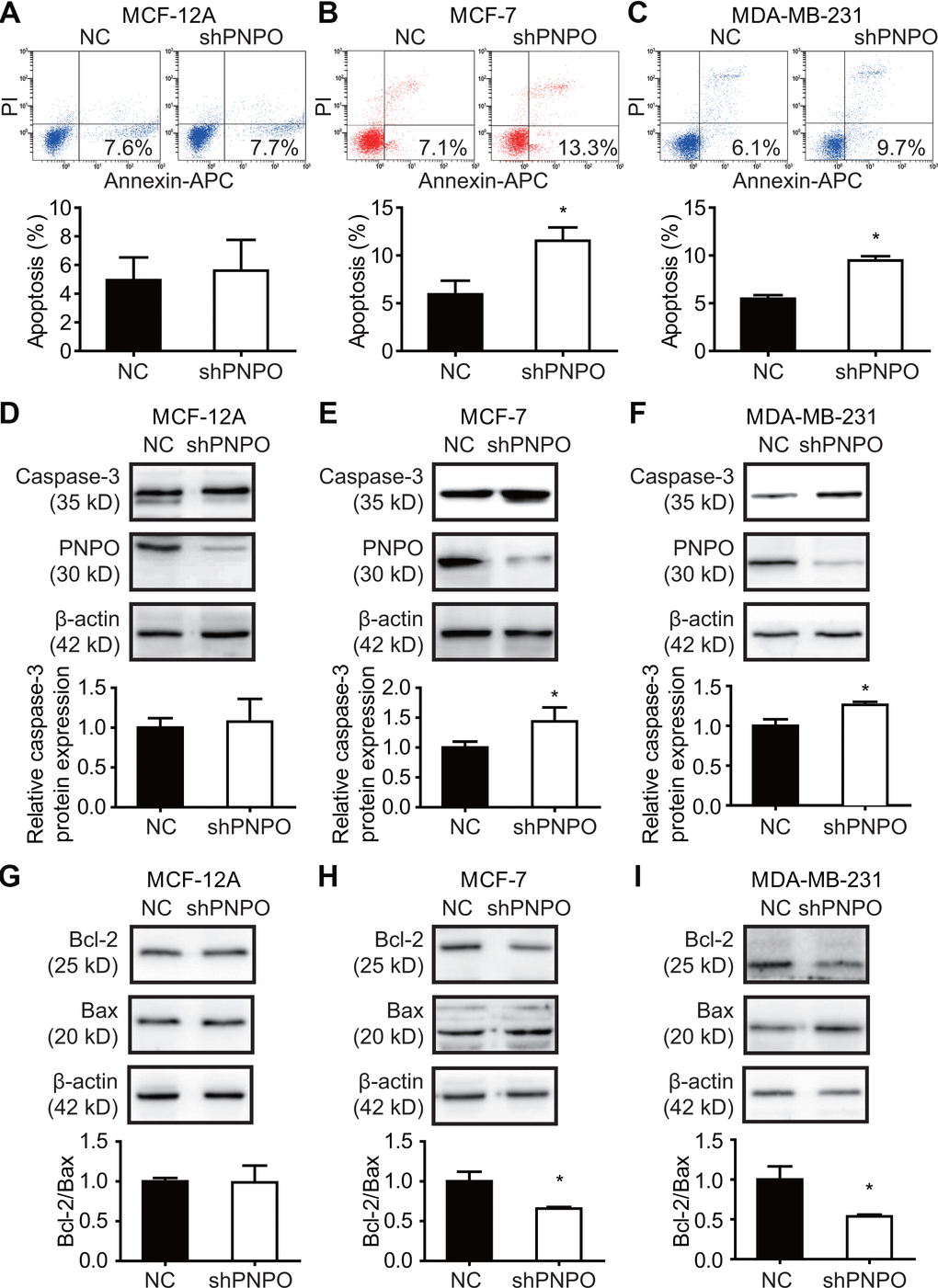
Figure 8.The effect of PNPO on breast cell apoptosis. (A–C) Apoptotic cells were detected by flow cytometry after PNPO knockdown in MCF-12A (A), MCF-7 (B), and MDA-MB-231 (C) cells. (D–F) Expression of caspase-3 and PNPO protein after PNPO knockdown in MCF-12A (D), MCF-7 (E), and MDA-MB-231 (F) cells detection by Western blot. (G–I) Expression of Bcl-2 and Bax protein after PNPO knockdown in MCF-12A (G), MCF-7 (H), and MDA-MB-231 (I) cells detection by Western blot. Histograms show the quantitative analyses. NC, negative control of shRNA; shPNPO, PNPO-shRNA. n =3; * P < 0.05.