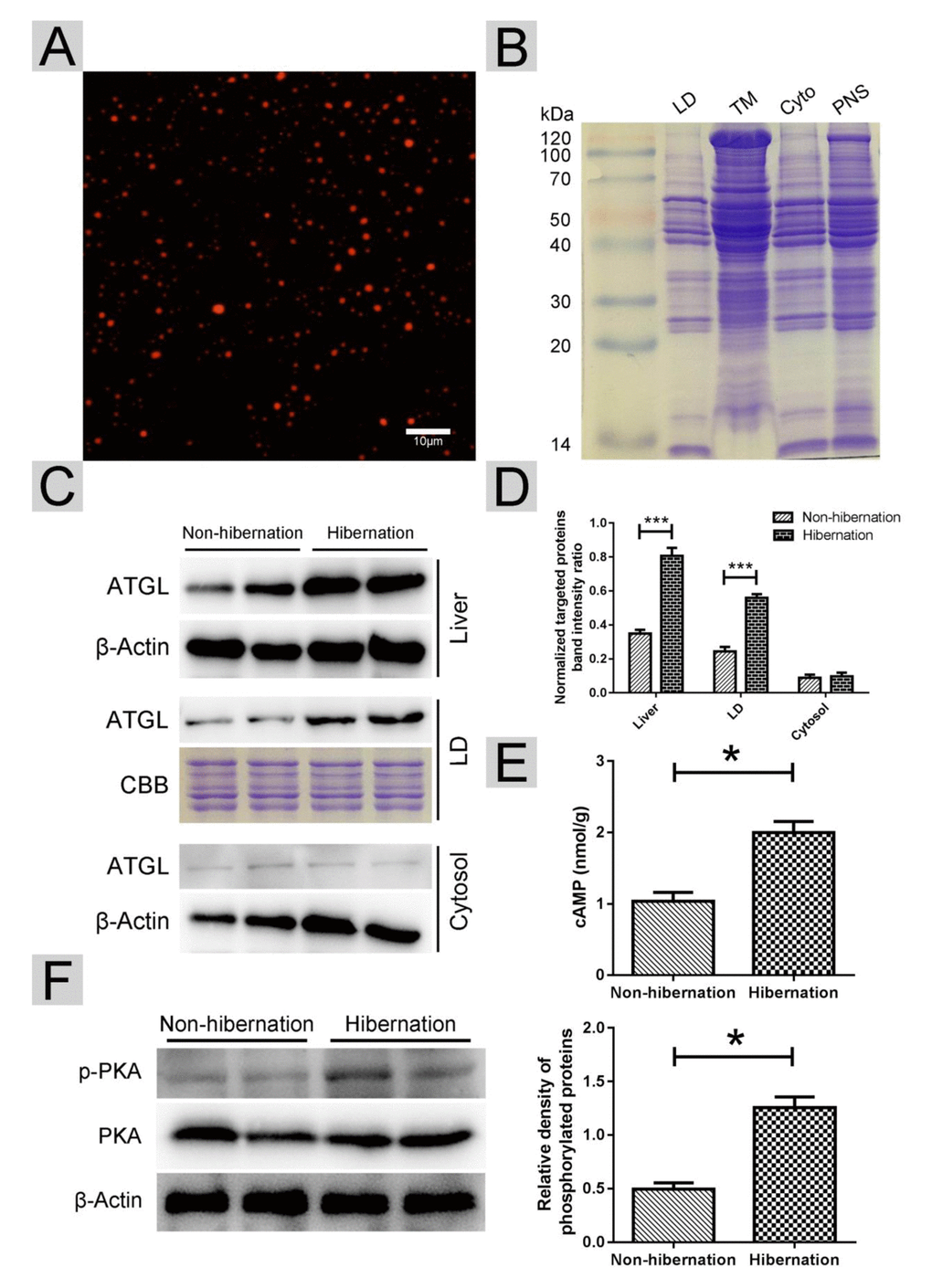
Figure 4.Activated cAMP/PKA signal increased ATGL accumulation on the surface of LDs. (A) Nile red staining of isolated LDs; (B) Colloidal blue staining of LDs, total membrane (TM), cytosol (Cyto) and post-nuclear supernatant (PNS) in the liver; (C) Western blot analysis of ATGL protein expression in the liver, LDs and cytosol; (D) Statistics of ATGL protein expression; (E) The cAMP level in the liver; (F) Differential phosphorylation of PKA in the liver.