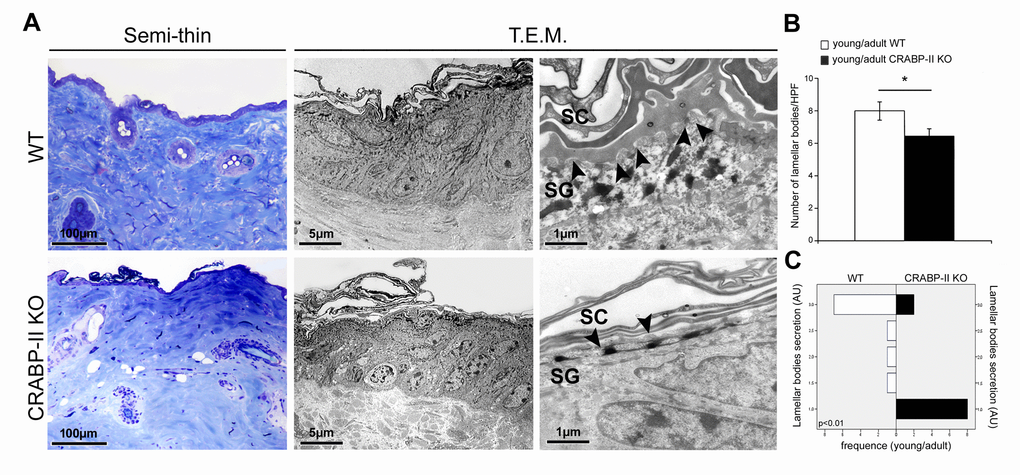
Figure 5.Ultrastructural evidence of age-related epidermal damage is early and greater in CRABP-II knock-out mice. (A) Representative images of toluidine blue-stained semithin EPON-embedded sections and transmission electron microscopy (T.E.M.) photographs of epidermis of young/adult wild-type (WT) and CRABP-II knock-out (KO) mice. (B) Bar graphs show the semiquantitative evaluation of ultrastructural epidermal number of lamellar bodies (n=10 young/adult WT and n=10 young/adult CRABP-II KO). Values are group mean ± SEM, t-Test: * indicates p< 0.05. (C) Semiquantitative evaluation of ultrastructural epidermal secretion of lamellar bodies (n=10 young/adult WT and n=10 young/adult CRABP-II KO). Mann-Whitney’s U-test. Arrow heads indicate lamellar bodies. Abbreviations: SC, stratum corneum; SG, stratum granulosum; HPF, High Power Field; AU, arbitrary units.