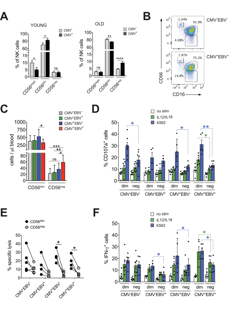
Figure 1.CD56neg NK cells with impaired effector function expand in CMV and EBV co-infected individuals >60 years of age. (A) Frequencies of CD56bright, CD56dim and CD56neg NK cells in YOUNG (<35 years) CMV– (gray bars, n=10/10) and CMV+ (black bars, n=10/10) individuals compared to OLD (>60 years) CMV- (gray bars, n=20/21) and CMV+ (black bars, n=17/20) donors analyzed as in Supplementary Figure S1A. (B) Representative FACS dot plots from a CMV–EBV– and a CMV+EBV+ donor are shown. Numbers indicate the percentage of cells within total NK cells in peripheral blood. (C) Absolute cell numbers for CD56dim and CD56neg NK cells – as determined by FACS analysis in total PBMCs– are shown in a cohort of HDs >60 years of age stratified as CMV–EBV– (n=11/11), CMV–EBV+ (n=10/24), CMV+EBV– (n=6/6), and CMV+EBV+ (n=12/14). (D-F) FACS-sorted CD56dim and CD56neg NK cells from CMV–EBV– (n=7), CMV–EBV+ (n=4), CMV+EBV- (n=4) and CMV+EBV+ (n=5) donors were either left un-stimulated (empty bars), stimulated with IL-12 / IL-18 (green bars) or K562 target cells (blue bars) and (D) CD107a expression (E) target cell lysis and (F) IFN-γ production were assessed after 6 hours of (co-)culture. Parametric data were compared by Student’s t-test and are shown as mean ± SEM, non-parametric data by Mann-Whitney test and are shown as median ± IQR, respectively. * p≤0.05, ** p≤0.005, *** p≤0.005.
Figure 1 — CD56-negative NK cells with impaired effector function expand in CMV and EBV co-infected healthy donors with age | Aging