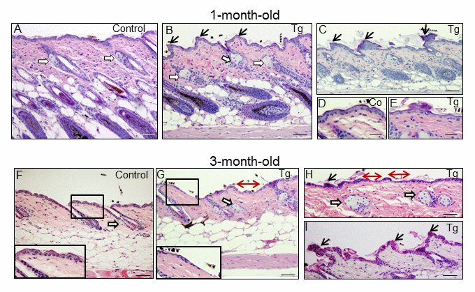
Figure 3.Histological alterations in the back skin of 1 and 3-month-old K5-CYLDC/S mice. (A-E) Histology of the back skin of 1-month-old Control (A, D) and K5-CYLDC/S mice (B, C, E). (A) Observe small sebaceous glands (white arrows) and HFs in the anagen phase in Control mice. (B, C) Note in transgenic mice the presence of moderately hyperplastic sebaceous glands, epidermal ridges and HFs initiating the anagen phase of the second hair growth cycle. (D, E) Slight thinning of the epidermis of K5-CYLDC/S mice. (F-I) Histology of the back skin of 3-month-old Control (F) and K5-CYLDC/S mice (G-I). (F) Note small sebaceous glands and telogenic HFs in Control mice. (G-I) Observe marked epidermal atrophy; abundant epidermal ridges of pyknotic keratinocytes, increased hyperplasia of the sebaceous glands and areas with orphan sebaceous glands lacking hair follicles in transgenic mice. White arrows: sebaceous glands; black arrows: epidermal ridges of pyknotic keratinocytes; double-headed red arrows: areas of epidermal atrophy. Scale bars: 250 μm (C, F-H); 200 μm (A, B); 180 μm (I); 150 μm (D, E).