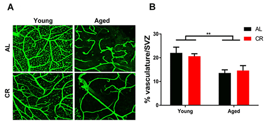
Figure 4.Vascular density declines in the aged subventricular zone despite dietary intervention. (A) Confocal images of the SVZ vascular plexus labeled with laminin. (B) Quantification of the vasculature density. ** = p< 0.01, two-way ANOVA, data are presented as mean ± SEM, n=5/group.