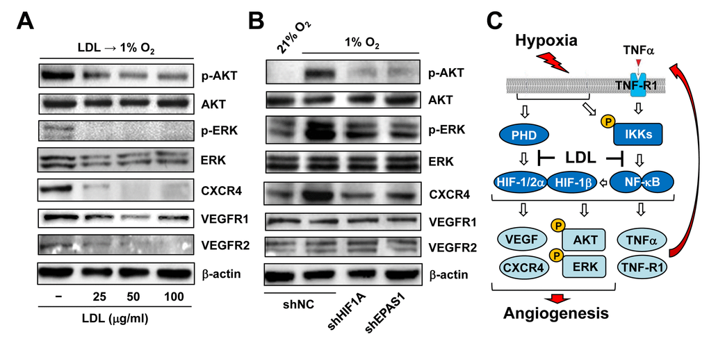
Figure 7.LDL-mediated impairment of hypoxia-induced angiogenesis involves a broad spectrum of HIF-dependent signaling pathways. (A) HUVECs were exposed to indicated concentrations of LDL (25 - 100 μg/ml) for 48 hrs, and then cultured under hypoxic (1% O2) condition for additional 48 hrs. (B) HUVECs with shRNA knockdown of HIF-1α or HIF-2α were exposed to 1% O2 (21% O2 as normoxia control) for 24 hrs. After treatment, Western blot analysis was performed to monitor phosphorylation of AKT (S473), ERK1/2 (T202/Y204), CXCR4, VEGFR1, and VEGFR2. Blots were reprobed for β-actin as loading control. (C) A schematic diagram for the mechanism by which hypoxia induces angiogenesis via an autocrine loop of TNFα and resulting activation of the self-regulatory TNFα/NF-κB/HIF/VEGF signaling network in ECs, as well as the potential mechanism of action for anti-angiogenic property of LDL by which a) LDL might impair the autocrine loop of TNFα via down-regulation of its receptor TNF-R1, rather than TNFα itself; and b) LDL disrupts the TNFα-NF-κB-HIF-VEGF signaling cascade via down-regulation of multiple key components of both canonical and non-canonical NF-κB pathways.