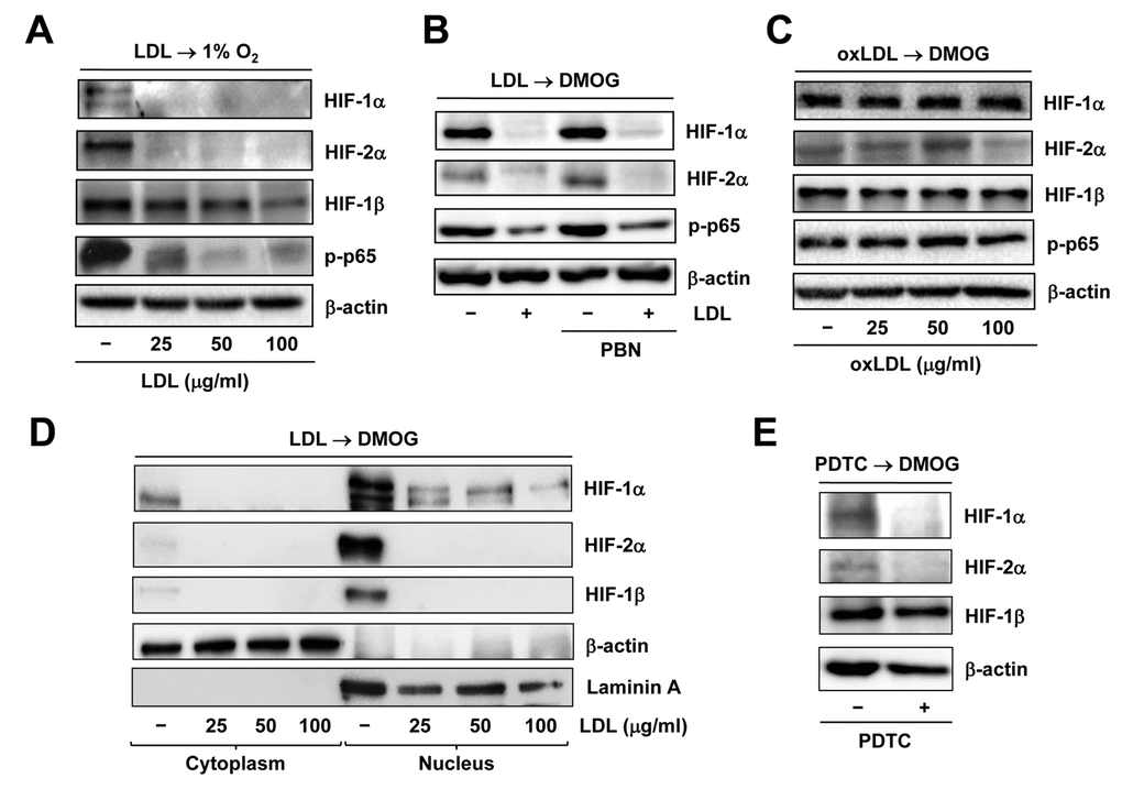
Figure 4.Native LDL, but not oxLDL, inhibits activation of HIF and NF−κB signals induced by hypoxia. HUVECs were treated as follows: (A) pre-treated with LDL (100 μg/ml) for 48 hrs, followed by incubation under hypoxic (1% O2) condition for additional 48 hrs; (B) pre-treated with LDL (100 μg/ml) in the presence or absence of the free radical scavenger PBN (2 mM) for 48 hrs, and then exposed to the PHD inhibitor DMOG (1 μM) for additional 72 hrs; (C) pre-treated with the indicated concentrations of oxidized LDL (oxLDL, μg/ml) for 48 hrs, followed by DMOG (1 μM) for additional 72 hrs. After treatment, Western blot analysis was performed to monitor expression of HIF-1α, HIF-2α, and HIF-1β, as well as phosphorylation of NF-κB p65 (S536). (D) Alternatively, HUVECs were treated as described in panel 4B, after which cytoplasmic and nuclear fractions were separated and subjected to Western blot analysis for monitoring nuclear translocation of HIF-1α, HIF-2α, and HIF-1β. Blots were reprobed for β-actin and laminin A as loading controls for cytoplasmic and nuclear fractions, respectively. (E) HUVECs were pre-incubated with the NF-κB inhibitor PDTC (100 μM) for 4 hrs, followed by DMOG (1 μM) for additional 72 hrs, after which the protein levels of HIFs were monitored by Western blot analysis.