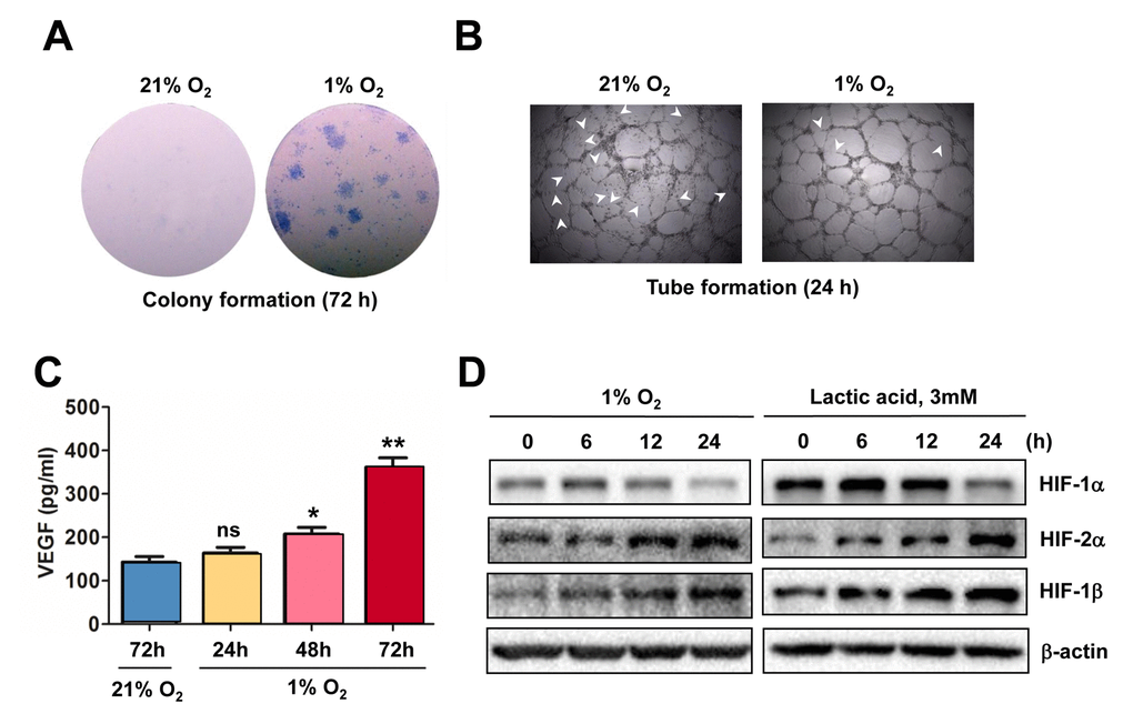
Figure 2.Hypoxia induces angiogenesis of ECs, in association with VEGF production and HIF activation. (A, B) HUVECs were cultured in ECGM medium containing 2% serum for the indicate intervals under either hypoxic (1% O2) or normoxic (21% O2) condition, after which cells were subjected to the following assays, including (A) colony formation assay (72 hrs) and (B) Matrigel-based tube formation assay (24 hrs). Representative microscopic images for at least three independent experiments were shown. Arrowheads indicate unclosed loops of vascular structure. (C) When HUVECs were cultured as described above under hypoxic (1% O2) or normoxic (21% O2) condition, medium was harvested at the indicated intervals and subjected to an ELISA assay to determine absolute amount of VEGF (pg/ml). Values represent the means ± SD for at least three independent experiments performed in triplicate. *P < 0.05 and **P < 0.01 for comparison with control (72 hrs under 21% O2); ns = not significant. (D) HUVECs were cultured under hypoxic (1% O2) condition (left panels) or exposed to the chemical hypoxia mimetic lactic acid (3 mM) for the indicated intervals (6 - 24 hrs), after which Western blot analysis was performed to monitor expression of HIF-1α, HIF-2α, and HIF-1β. Blots were reprobed for β−actin as loading control.