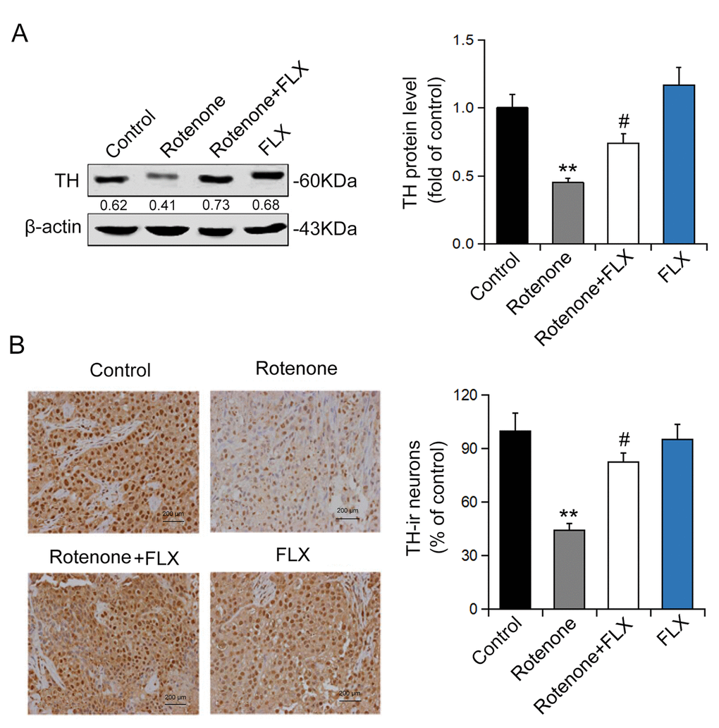
Figure 3.Effects of FLX on DA neurons loss in PD rats. (A) TH protein expression was detected using Western blot, n=6. (B) TH-positive DA neurons were determined using immunohistochemistry. The ratio of TH-positive neurons in experimental groups to those cells in the control group was evaluated. n=4, **P<0.01 versus control group; #P<0.05 versus rotenone-treated group.