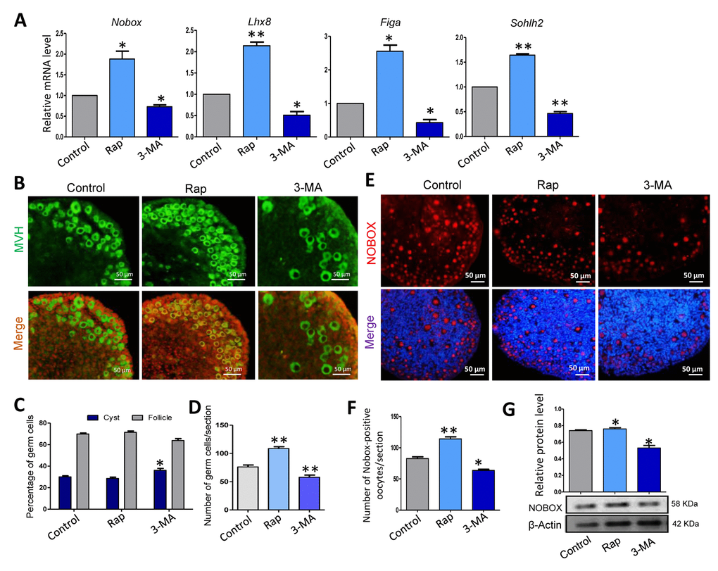
Figure 6.Rapamycin-promoted autophagy and prevented germ cell over loss after 5 days of treatment. (A) Quantitative RT-PCR for Nobox, Lhx8, Figa and Sohlh2 mRNA levels in control, rapamycin and 3-MA treated ovaries for 5 days. (B) IF staining for MVH (green) of control, rapamycin and 3-MA treated mouse ovaries for 5 days. (C) Percentage of germ cells in cysts and follicles in the three groups after 5 days treatment. (D) Average number of survived oocytes in the three groups. Autophagy leads to more survival of gem cells. (E-F) Number of NOBOX-positive oocytes/section in control, rapamycin and 3-MA treated ovaries. (G) Level of NOBOX protein in control, rapamycin and 3-MA treated ovaries. The results are presented as mean ± SD. * P < 0.05; ** P < 0.01.