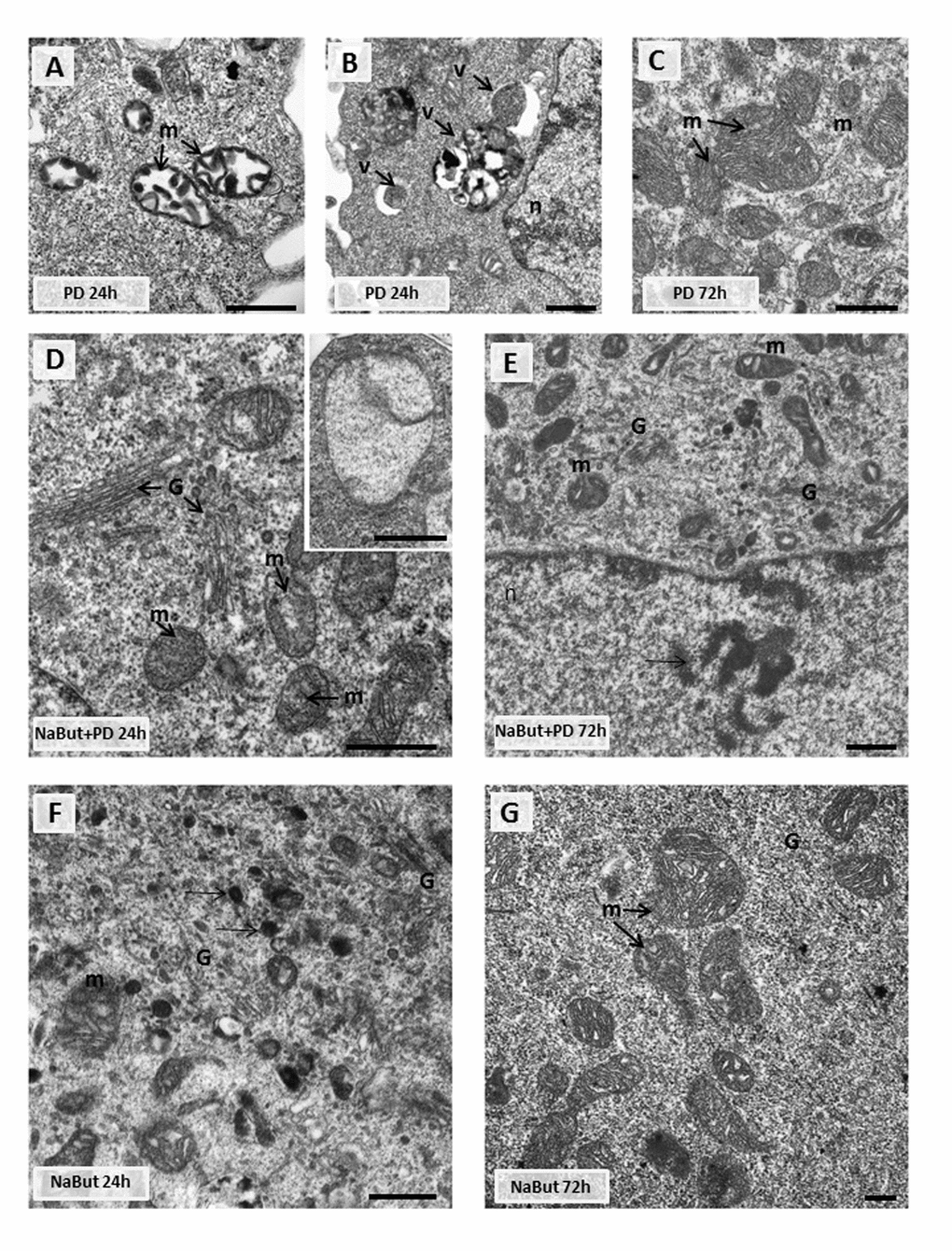
Figure 4.Transmission electron microscopy (TEM) images showing the ultrastructure of intact and senescent ERas cells treated with inhibitor of MEK/ERK-pathway (A,B,C). Representative images of the mitochondria in intact ERas cells after MEK/ERK suppression. Note severe alterations of mitochondria (m) after 24 hours of treatment (A), mitochondria in autophagosome-like vesicles (B) and normal-looking mitochondria after 72 hours of treatment (C). (D) Representative image of senescent ERas cell treated with PD for 24 hours. It contains well-developed Golgi apparatus (G) and mitochondria (m) with rare cristae. Inset: mitochondria with complete loss of the cristae and the preserved double-membrane envelope. (E) Representative image of senescent ERas cell treated with PD for 72 hours. The cell contains well-developed Golgi apparatus and swollen mitochondria with vacuolar structure. Note poorly developed nucleoli (arrow). The senescent ERas cells after 24 (F) and 72 (G) hours of NaBut exposure. Stacks of Golgi cisternae and lysosome-like structures (F, arrows) can be seen. Designations: G, Golgi; m, mitochondria; n, nucleus; v, vesicle. Scale bars: 500 nm.