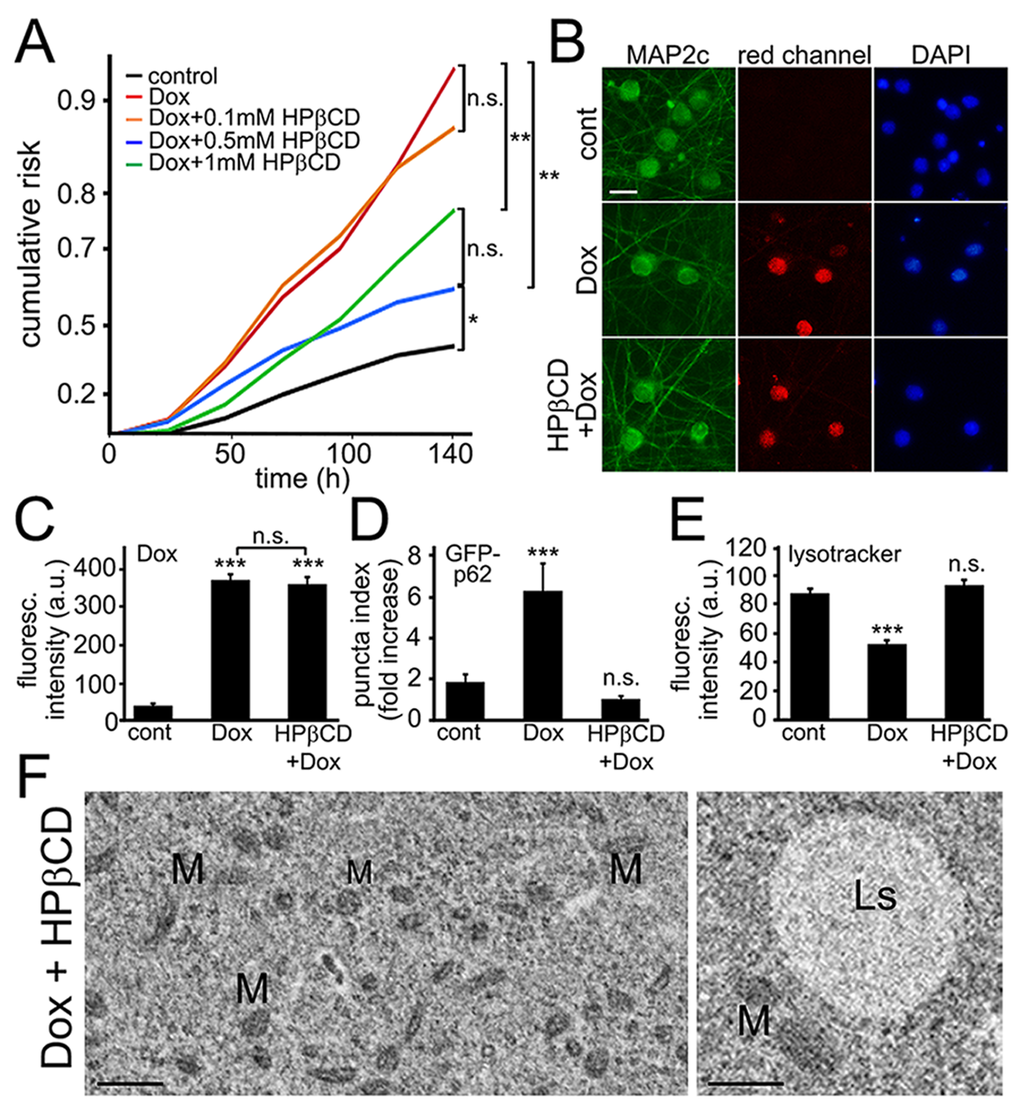
Figure 6.Cyclodextrin reduces neuronal damage induced by exposure to doxorubicin. (A) Survival analysis of five neuronal cohorts transfected with mApple (a morphology and viability marker) were treated with a vehicle or with doxorubicin alone or in combination with HPβCD. The first neuronal cohort was treated with a vehicle. The second neuronal cohort was treated with 10 nM doxorubicin. The remaining three neuronal cohorts were treated with 10 nM doxorubicin in combination with 0.1 mM HPβCD (third neuronal cohort), 10 nM doxorubicin and 0.5 mM HPβCD (fourth neuronal cohort), and 10 nM doxorubicin and 1 mM HPβCD (fifth neuronal cohort). Neurons were tracked and imaged over 6 days. Risk of death associated with a treatment for each cohort was calculated with JMP software. *p<0.01, **p < 0.001 (Log-Rank test), n.s., non-significant. One hundred fifty neurons were analyzed per condition. Results were pooled from two independent experiments. (B) HPβCD does not prevent doxorubicin from binding to DNA. Cortical neurons were treated with 50 nM doxorubicin or with 50 nM doxorubicin and 0.5 mM HPβCD overnight, fixed, and stained for MAP2c (green) and with the nuclear Hoechst dye (blue), and imaged. Scale bar is 20 μm. (C) Images of fixed neurons from (B) were analyzed. ***p<0.0001 (t-test), n.s., non-significant. Three hundred neurons were analyzed per condition. Results were pooled from two independent experiments. (D) Three cohorts of cortical neurons were transfected with mApple and GFP-p62. The first neuronal cohort was treated overnight with a vehicle, the second cohort was treated with 50 nM doxorubicin (overnight), and the third cohort was co-treated with doxorubicin and 0.5 mM HPβCD (overnight). Quantification of fluorescent images revealed that HPβCD lowers the levels of GFP-p62 in doxorubicin-treated neurons. ***p < 0.0001 (t-test), n.s., non-significant. Two hundred neurons were analyzed. Results were from three independent experiments. (E) Cortical neurons were treated overnight with a vehicle or 50 nM doxorubicin or with a combination of doxorubicin and 0.5 mM HPβCD, and stained with a green lysotracker dye. HPβCD lowers the pH in in doxorubicin-treated neurons. ***p < 0.0001 (t-test), n.s., non-significant. Two hundred neurons were analyzed. Results were pooled from three independent experiments. (F) Electron micrographs of cultured cortical neurons co-treated with doxorubicin and 0.5 mM HPβCD (overnight). Left panel: Note that the cytoplasm lack the damage observed in neurons treated with doxorubicin alone. M, mitochondria. Bar, 1 µm. Right panel: Ls, lysosome; M, mitochondria. Bar, 500 nm.