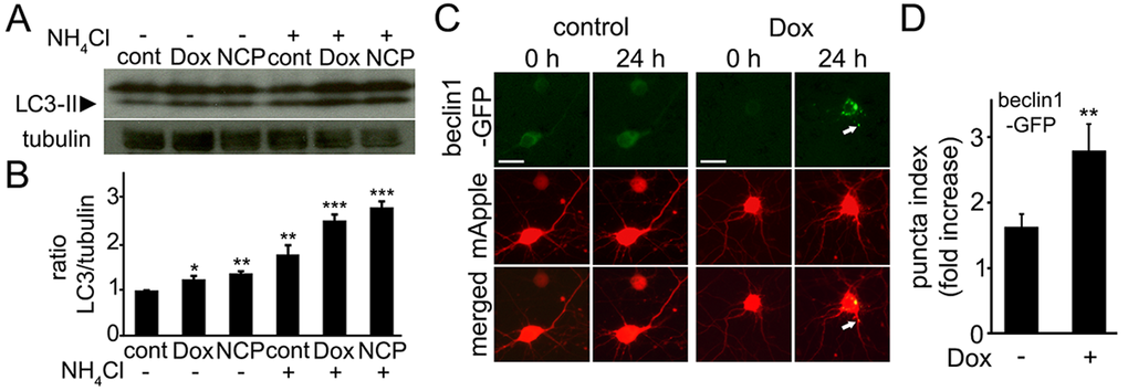
Figure 1.Doxorubicin induces autophagy in primary cortical neurons. (A) Autophagy is induced in cultured primary cortical neurons by 50 nM doxorubicin (overnight) as reflected by the increased levels of LC3-II. Tubulin was used as a loading control. LC3-II accumulated in neurons treated overnight with 50 nM doxorubicin or 5 µM 10-NCP (an autophagy enhancer as positive control) with or without 10 mM NH4Cl, 4 h. LC3-II increased in doxorubicin-treated cells when NH4Cl was added reflecting enhanced autophagic flux. (B) Measurements of the LC3-II bands from (A). The LC3-II intensities were normalized to the tubulin loading control. *p<0.01, **p<0.001, ***p<0.0001 (ANOVA). Results were pooled from three independent experiments. (C) Doxorubicin promotes the formation of pre-autophagosomal complexes as reflected by beclin1-GFP-positive puncta. Cortical neurons were transfected with mApple (a morphology and viability marker) and beclin1-GFP, and treated with a vehicle or 50 nM doxorubicin (overnight). Note changes in beclin1-GFP localization, consistent with beclin1 relocalization to pre-autophagosomal structures. White arrow points a beclin1-GFP-positive structure in a neurite. Bar, 20 μm. (D) To score autophagy induction, the redistribution of beclin1-GFP into puncta, is reflected by the fold-increase of puncta index, which is the standard deviation among pixels within the cellular region of interest. The puncta index significantly increased in doxorubicin-treated neurons. **p<0.001 (t-test). Two hundred neurons were analyzed from two independent experiments.