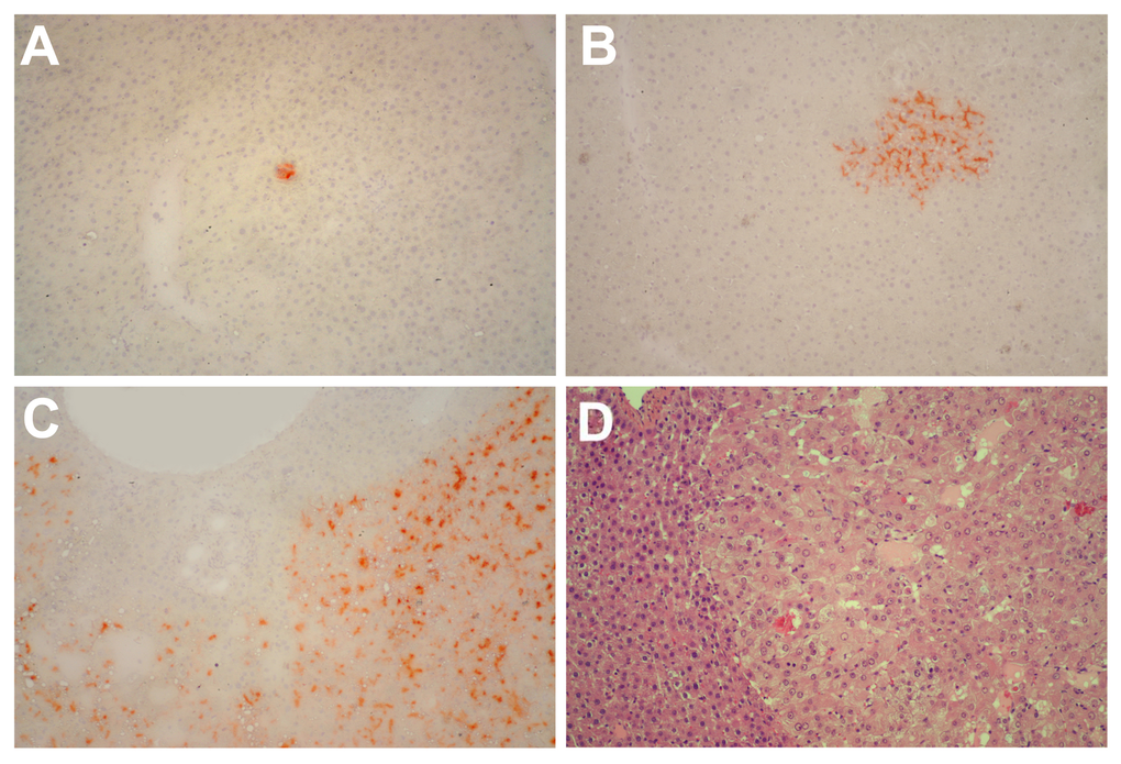
Figure 3.Panels (A) and (B) Histochemical staining for DPPIV of liver samples obtained from rats transplanted with pre-neoplastic hepatocytes at young age and killed 8 months after Tx. Small clusters of DPPIV-expressing hepatocytes (orange-rust) were discerned in all animals. Histochemical staining for DPPIV (panel C) and standard H&E staining (panel D) of a large hepatic nodule from a rat transplanted with pre-neoplastic hepatocytes at old age and killed 8 months later. Note the typical appearance of nodular hepatocytes arranged in multiple cell-thick plates. Magnification: 100x.