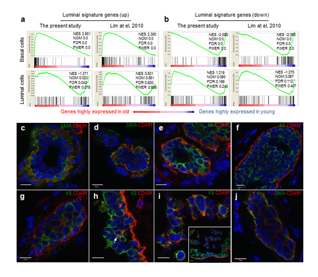
Figure 3.Aged basal cells were enriched with cells expressing luminal gene signature and contains K8+ luminal cells. (a and b) Gene set enrichment analysis. Genes that are expressed in at least one sample (normalized number of reads > 1) were rank ordered according to their fold changes between young (4-6 mon.) and old (30-32 mon.), with genes highly expressed in old cells on the left. Two sets (the present study and that of Lim et al., 2010) of luminal signature genes were analyzed and indicated as black bars in the plots. The luminal cell signature genes were significantly enriched in the old basal cells, and no significant enrichment was seen in young or old luminal cells. NES, normalized enrichment score. NOM, nominal p-value. FDR, false discovery rate. FEWR, familywise error rate p-value. (c-j) Immunostaining of representative mammary ducts from old (26-31 mon., n = 6) C57BL6/J mice showing that basal cells were CD49fhi, SMA+ and K8- in the majority normal (c, e) and hyperplastic ducts (d, f), but the presence of CD49fhi luminal cells (K8+, SMA-) in a few hyperplastic lesions (g-j). The inset in Panel i shows dislodged cells from the ductal wall at a lower magnification than the main panel. Scale bars, 10 μm.