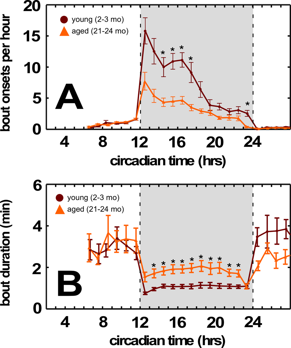
Figure 3.Aged BALB mice have fewer feeding bouts during the dark cycle. (A) Decreased feeding bouts in aged BALB mice. (B) Increased feeding bout duration in aged BALB mice. Traces in light orange correspond to young mice; traces in dark orange correspond to aged mice. Grayed region depicts dark cycle, dashed lines indicate dark cycle onset and offset, respectively. Asterisks indicate p<0.01, Bonferroni corrected; error bars are ± 1 standard error of the mean.