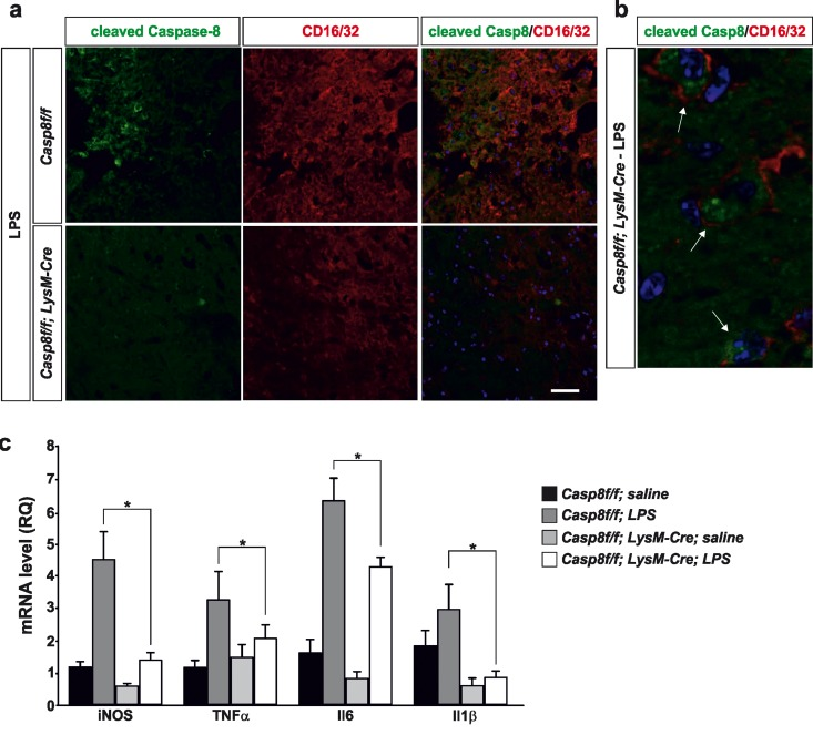
Figure 3.Caspase-8 is activated in viable CD16/32-immunolabeled microglia in substantia nigra in response to intranigral LPS injection, which is associated to higher key proinflammatory microglial markersPanel (a) shows an illustration of dual immunofluorescence of cleaved caspase-8 and CD16/32 in substantia nigra in response to intranigral LPS in Casp8fl/fl mice and CreLysMCasp8fl/fl mice. Note how cleaved caspase-8 labeling is mostly associated to CD16/32-labeled microglia, which is dramatically decreased in CreLysMCasp8fl/fl mice. Panel (b) shows higher magnification photograph of that shown in panel (a) demonstrating active caspase-8 within viable CD16/32-immunolabeled microglia in Casp8fl/fl mice after LPS. Panel (c) shows the effect of LPS on mRNA expression of inducible nitric oxide synthase (iNOS), tumour necrosis factor α (TNF- α), interleukin 6 (IL-6) and interleukin-1β in substantia nigra of Casp8fl/fl mice and CreLysMCasp8fl/fl mice. mRNA levels were measured by quantitative PCR. Results are mean ± SD of at least four independent experiments and are expressed as relative quantification (RQ), calculated using the delta Ct method. Statistical significance was calculated by one-way analysis of variance followed by the least significant difference post hoc test for multiple range (p <0.05). As expected, LPS injection increased the expression levels of mRNA in Casp8fl/fl mice (WT). This induction was highly prevented in CreLysMCasp8fl/fl mice (KO). Note the extremely low levels of IL-1β in CreLysMCasp8fl/fl mice. Scale bar: a: 30 μm; b: 10 μm.