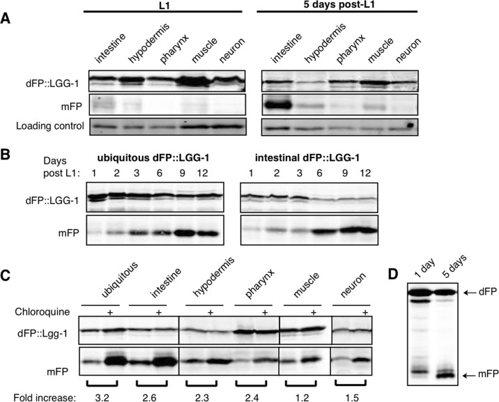
Figure 7.Autophagic flux increases with age(A) L1 animals have low levels of mFP in all tissues (left panel), while animals aged to five days after L1 show an increase in mFP in most tissues (right panel, quantifications shown in Supplementary Table 1). Representative blots (of n=4 experiments) are shown. (B) Animals expressing either ubiquitous or intestinal dFP::LGG-1 show a shift in autophagy that occurs in early adulthood, with a dramatic increase in the amount of mFP accumulation between three and six days of life. (C) Animals aged five days and treated with chloroquine for 18 hours accumulate more mFP in all tissues, showing that mFP accumulation is not due to a dramatic change in lysosome function. All lanes are from the same experiment run in the same SDS-PAGE gel, with delineations indicating areas of the image that were separately adjusted for brightness and contrast for the sake of clarity (uncropped immunoblots are shown in Supplementary Figure 1). (D) Release of mFP from soluble dFP reporter also shows a shift in mFP accumulation with age, with increasing amounts of mFP at five days of age.