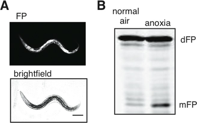
Figure 5.Cytoplasmic flux through the autophagic pathway(A) Ubiquitously-expressed soluble dFP was present in all tissues, as visualized with fluorescence. Scale bar 100mm. (B) This reporter showed a substantial increase in mFP release when the nematodes were exposed to anoxia. This demonstrates an increase of bulk autophagic flux without overexpression of any protein involved in autophagy.